
The mind-mucus connection
When phlegm runs amok, it can be life-threatening. Neuroscience know-how offers a way to put a cork in it.
Next time it feels like you’re sneezing your brains out or coughing up a lung, consider that brains and lungs have something very much in common: They share some secrets about secretion. Nerve cells in the brain take the high road, emitting bursts of chemicals in order to pass their signals from one to the next (a process known as neurotransmission). Goblet cells in the lung take the low road, squirting out rivers of mucus when they get irritated.
Yep, mucus. Like it or lump it, we can’t live without it. We don’t think too much about the sometimes slimy, sometimes sticky, sometimes lumpy stuff, and when we do, we don’t think much of it. But our health hinges on it.
In the right amounts, at the right consistency, mucus is a lung’s best friend. It’s also essential to the proper function of the stomach, intestine and urogenital tract. But if there’s too much of it, or if it’s too adhesive, it can betray the organ it is meant to serve.
Medications for chronic obstructive pulmonary disease, which also affects about 25 million in the United States, are even less likely to work. Excess mucus also spells trouble for the 1 in 20 people who develop acute bronchitis each year. And for the roughly 30,000 Americans who have cystic fibrosis.
Oddly, the excessive buildup of mucus in the lungs and airways bears some powerful resemblance, at the molecular level, to the way nerve cells in the brain secrete pulses of specialized chemicals to transmit signals to one another. In a sense, these chemicals, called neurotransmitters, form the substrate of our soul: They guide our every cognition, emotion, motion and ambition.
To carry out their lofty job description, neurotransmitters need to be released in a precise manner. Mucus, not so much — “precision mucus” is not a thing. But there’s still a big difference, healthwise, between just enough of it and way too much of it.
Any resemblances between neurotransmission and mucus hypersecretion do not extend to the attention they get from researchers. Walk the halls of any solid medical school, and you will come across departments of neurology, neurobiology, neurosurgery and psychiatry. If you were to stumble on a department of mucus, your first instinct would probably be to pick up your pace or to attempt to wake up.
It stands to reason that the obvious importance of a functioning nervous system would result in quite a lot of research attention being focused on neurotransmission. Mucus secretion, although vitally important, is less glamorous, far more esoteric, and more quietly explored.
But the burden of medical disorders caused or exacerbated by too much mucus is nothing to sneeze at. As fate would have it, hard-won insights from the world of neuroscience are now directing beams of understanding at lung disorders caused by too much mucus.
Connections between mucus and the brain
Using techniques originally designed to tease apart the functions of several proteins that work together to coordinate the release of neurotransmitters, a team including a neurotransmission expert and a mucus explorer has measured and modified the workings of the pathway responsible for excessive release of a key protein in mucus. This may soon pay off in the form of entirely new, precisely targeted treatments for mucus-stressed lungs.
The leader of the project, Axel Brunger, PhD, a Stanford Medicine professor of molecular and cellular physiology, of neurology and neurological sciences and of photon science, has a history of research delineating the workings of neurotransmission.
In the course of his career, he has collaborated frequently with Tom Südhof, MD, a professor of molecular and cellular physiology who received a Nobel Prize for demonstrating how myriad tiny bubble-like packets, or vesicles, containing neurotransmitters are held in just the right place inside a nerve cell, ready to release the signal-bearing molecules at just the right moment.
Armed with this understanding, Brunger, other Stanford Medicine investigators and collaborators at the University of Texas MD Anderson Cancer Center in Houston and at Ulm University in Germany have designed a compound that’s capable of blocking mucus hypersecretion while, crucially, not interfering with the necessary low-level secretion of the gummy substance.
“This is the first compound that specifically alleviates the pathological hypersecretion of mucus common to cystic fibrosis, chronic obstructive pulmonary disease, asthma, viral respiratory infections and more,” said Brunger, who is a Howard Hughes Medical Institute investigator.
The discovery, described in a study published in March in Nature, could improve the lives of millions who suffer from airway obstruction caused by excess mucus.
What we call breakthroughs seldom come about by a burst of insight. They more often unfold inch by painstaking experimental inch, sometimes accelerated by the serendipitous intersection of two minds.
“This is the first compound that specifically alleviates the pathological hypersecretion of mucus common to cystic fibrosis, chronic obstructive pulmonary disease, asthma, viral respiratory infections and more.”
Axel Brunger, PhD, a professor of molecular and cellular physiology, of neurology and neurological sciences and of photon science
In landmark papers published in the past 25 years, Brunger and various co-authors (including Südhof) discovered the myriad molecular details of how a select group of proteins cooperate to orchestrate neurotransmission.
Burton Dickey, MD, a mucus expert and professor of pulmonary medicine at the MD Anderson Cancer Center, has in years past collaborated with Südhof on neurotransmission research.
Work by Dickey and other scientists has shown that upsized secretion of mucin — a long, stringy protein that’s mucus’s distinguishing component — works in much the same way as neurotransmission, involving analogous vesicles and collaborating proteins. One crucial difference: The vesicles are filled with mucin instead of neurotransmitters.
To advance his neurotransmission research, Brunger invented a technique that enables him to observe how adding selected proteins to stripped-down laboratory models of single vesicles affect a vesicle’s behavior — for example, in relation to a similarly stripped-down and tweaked version of a cell’s outer membrane. This method would come in handy in unexpected ways.
In May 2016, Brunger gave a lecture at the Baylor College of Medicine in Houston. In the audience sat Dickey, who had heard about Brunger’s work from Südhof, a professor at the University of Texas before coming to Stanford.
Dickey approached Brunger after the lecture, told him he wanted to develop a compound to selectively block mucin hypersecretion — that is, to stop it in its tracks without interfering with the constant low-key output of mucin that ensures adequate mucus levels for proper organ function — and asked him if he’d care to collaborate. They became co-senior authors of the study.
Mucus facts
Mucus may not get talked about much at swank soirees, but there it is, lurking inside of every guest. It comes in various colors and viscosities, and hails from various regions of the body besides the lungs. There’s plenty of it in the nose. Saliva is mostly mucus. You also find it in the stomach and gut. (Mucus expelled from the lung is known as phlegm, or sputum.)
Different microbes impart different colors to mucus. Some molds are black, and they can turn mucus black. Blood makes it dark red.
But superficial differences aside, there are two main ingredients.
A healthy respiratory system’s mucus is 97% water. The other main ingredient, mucin, is secreted by goblet cells and seromucous glands at or just below the surface, or epithelium, of the nose, throat, bronchial tubes and small airways in the lungs. Lengthy chains of sugar molecules sprout from each mucin molecule’s surface, predisposing the protein to absorb water.
Mucin molecules readily cross-link into networks, forming the viscous gel we know so well. Secreted at moderate levels, mucus coats airway surfaces, serving as a lubricant and a protective barrier as well as a straitjacket for encapsulating microbial pathogens. The encased microbes are driven out of the lungs and upward in the airways by hairlike structures called cilia that project from cells abundant in the airway lining.
Few of us spend any time pondering these minutiae because a steady hum of crucial mucus secretion goes on pretty much all the time. We hardly notice it — until a fly lands in the ointment. Or until a microbe lands in the lungs.
Inflammation, often elicited by microbial pathogens, shifts mucin secretion into overdrive. That’s great for trapping and washing away offending microbes, but persistent inflammation can cause trouble.
What’s more, the newly secreted inflammation-triggered bolus of mucus often contains a higher-than-normal mucin content, increasing the liquid’s viscosity.
The thickened mucus can congeal into rubbery pads, or plaques, stifling gas exchange in the lungs and impeding airway cilia’s upward pumping of mucus-entrapped pathogens.
Secretion secrets
Evolution, having devised a nifty trick, would be remiss to relegate its execution to a single instance or an individual organ. Secretion of neurotransmitter chemicals in the brain and hypersecretion of mucin in the lungs work similarly.
In fact, some scientists think neurotransmission and mucus secretion may have evolved from the same ancient pathway — it’s also found in glue-secreting cells studding the tentacles the comb jelly, a jellyfish cousin, uses to capture plankton.
As we’ve seen, big bursts of mucin secretion and subsequent mucus buildup are driven by abnormal events such as inflammation. In the brain, analogous bursts of high-level secretion, far from being outliers, are critical to nerve cells’ normal function: signaling.
Neurotransmission — the relaying of impulses from one nerve cell, or neuron, to the next — depends on the carefully timed secretion of neurotransmitters from neurons’ tips. Those chemicals are routinely caged inside tiny vesicles situated near a neuron’s surface membrane like containers on a ship, waiting to be unloaded.
Spontaneous merging of these vesicles’ walls with the neuron’s outer membrane happens all the time, resulting in a fairly constant, low-level expulsion of the vesicles’ stored contents from the neuron. But electrical impulses traveling through the neuron trigger an abrupt, temporary influx of calcium into the neuron.
When calcium binds to a protein called synaptotagmin on the vesicles’ surfaces, synaptotagmin teams up with other proteins to tug some of the vesicles, winch-style, closer and closer to the neuron’s surface membrane until their enclosing membranes fuse with it, spilling the vesicles’ contents into the surrounding environment. The chemicals can then diffuse to nearby neurons, exciting or inhibiting activity in those recipient cells.
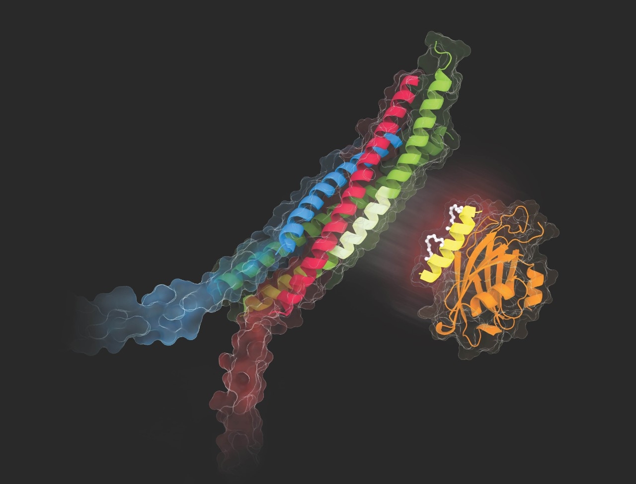
A closer look at synaptotagmin
In the new study, Brunger’s team showed that synaptotagmin is also essential to the rapid large-scale fusion of mucin-containing vesicles with secretory cells’ outer membrane — and to the resulting massive mucin expulsion.
And in studies in mice, they found that eliminating synaptotagmin dialed down mucin release considerably in inflammatory conditions that typically generate buckets of it, along with allergic reactions. If reducing the presence of synaptotagmin had this effect, then blocking its activity was clearly an approach worth exploring.
Brunger’s and Dickey’s groups designed a small protein snippet, or peptide, called SP9, that blocked synaptotagmin’s calcium-triggered interaction with its cooperating proteins in mice. SP9 is essentially a synthesized segment of a protein to which synaptotagmin binds. To stabilize SP9’s shape and ensure its effectiveness, the team bolstered the peptide with biochemical braces.
The study’s lead author, Ying Lai, PhD, then a postdoctoral scholar in Brunger’s lab, applied Brunger’s single-vesicle-modeling technique to mucin-containing vesicles and mucin-secreting cells’ outer membranes — the classic Brunger approach. In this simplified system, SP9 preferentially inhibited inflammation-driven hypersecretion of mucin, mucus’s chief protein constituent.
But the researchers needed to find a way for SP9 to transit the secretory cell’s outer membrane and get inside, where the action is. Working with Manfred Frick, PhD, a professor of medicine at Ulm University who became the study’s third co-senior author, Lai solved the problem by splicing SP9 to yet another peptide, PEN, that’s known to be excellent at penetrating cell membranes.
Frick’s group confirmed, in human cells cultured from lung-tissue biopsies, that the conjoined-peptide pair penetrated the secretory cells and blocked their inflammation-induced mucin secretion.
Dickey’s lab at MD Anderson then showed that PEN-SP9 administration in mice not only stymies inflammation-induced mucus secretion but also substantially reduces the plaque-plugged area in the mice’s lungs. That suggests it could be effective as a therapy in humans.
Toward clinical trials
Brunger and Dickey have filed a joint provisional patent application with Stanford’s Office of Technology Licensing and the MD Anderson Cancer Center’s Office of Technology Commercialization. Brunger hopes to see an optimized version of SP9 begin testing in patients within the next two to three years.
“This didn’t happen by accident,” Brunger said. “It’s been a long road with many failures, and it shows the importance of basic research — you just keep putting one foot in front of the other, and then suddenly you have something.”
