
Practice doesn’t always make perfect
Seizures worsen by co-opting one of the brain’s mechanisms for learning

Remember learning a tricky physical skill when you were a kid? Maybe you wobbled down the driveway on a two-wheeler, terrified of tipping over. Perhaps you spent your first few days of “knitting” poking yarn into frustrating snarls. Maybe you made a jangly racket as you lurched through your first two-handed piano scales.
You may have felt discombobulated, but, with repetition, things began to click. The bicycle stayed upright, the knitted and purled stitches slipped smoothly past your fingers, the notes sounded musical.
The skill became easier because your brain changed.
As you practiced, neurons choreographing the task sent signals not just to each other but also to nearby cells on the brain’s maintenance crew. They told those maintenance workers, “Help us out here. We need more insulation on these wires.”
Gradually, your cycling, knitting or piano-playing neurons — coated in more layers of insulating myelin, the fatty substance that gives the brain’s white matter its color — could send faster, better-coordinated signals. With your improved neurons, you could learn things that would once have seemed unthinkable: how to ride with no hands, execute a complicated cable stitch, play Für Elise.
The details of this process, called adaptive myelination, were first explored a decade ago by Stanford Medicine experts, led by Michelle Monje, MD, PhD, now the Milan Gambhir Professor in Pediatric Neuro-Oncology. Her team’s discoveries opened an exciting scientific vista. Adaptive myelination, which changes nerve signaling by modifying neuronal fibers’ insulation, represented a previously unappreciated mechanism for learning.
While scientists had long understood that learning changes the number and strength of connections between our neurons, adaptive myelination was recognized as a whole new way that experience could change us — that knowledge could become cemented in our brains.
But this mode of practice making perfect can also be hijacked by disease. A team of Stanford Medicine scientists, led by one of Monje’s former trainees, Juliet Knowles, MD, PhD, has recently shown that the brain can use adaptive myelination to perfect “skills” that are actually pathological, such as having seizures. The discovery has opened the way for investigations into whether drugs that target the unhelpful form of myelination could stop epilepsy from progressing.

The anatomy of a seizing brain
“There’s been this adage for a long time that ‘seizures beget seizures,’” said Knowles, now an assistant professor of neurology and of pediatrics at Stanford Medicine. “Untreated, the natural course of many forms of epilepsy is that seizures just become more frequent and severe.”
Pediatric epilepsy isn’t a monolith: The disease definition encompasses any lasting predisposition to seizures. It can range in severity and has many possible origins, including single-gene defects, combinations of genetic abnormalities or injuries that alter the brain’s structure.
“It’s an incredibly difficult disease for children and their families,” said Knowles, a pediatric neurologist who treats children with epilepsy at Stanford Medicine Children’s Health. “Seizures can happen at any minute. They leave affected children and families with a difficult sense of vulnerability and loss of control.”
Epilepsy symptoms can also include cognitive impairment, poor sleep and a range of other neuropsychiatric problems. And seizures come in a variety of forms: They can start from one point (focal) or engulf the whole brain at once (generalized). In the category of generalized seizures are several subtypes, from those that cause pronounced convulsions to so-called absence seizures so subtle they may go undetected for months.
“Seizures can happen at any minute. They leave affected children and families with a difficult sense of vulnerability and loss of control.”
Juliet Knowles, MD, PhD
What’s happening in the brain when someone seizes? “Normally, our brains are perceiving our environment, responding to it, figuring out when it’s time to eat, time to sleep and so on,” said John Huguenard, PhD, professor of neurology and of neurosurgery, who, with Monje, mentored Knowles’ postdoctoral work and now collaborates on Knowles’ research. “That process is very well organized. A seizure is a co-opting of the process, where the same neurons that normally perform cognition start to coordinate their activity in an unuseful way.”
As an absence seizure begins, the neurons involved begin to fire together — “they start listening to each other rather than the outside world,” Huguenard said. “Pretty soon they’ve all been recruited, all asked to join this other activity.” After a few seconds of abnormally synchronous firing, the seizure ends and normal cognition resumes. This pattern can repeat dozens or hundreds of times per day.
Preventing seizures in people with epilepsy is not easy. Finding the right medication usually takes trial and error, if it succeeds at all; many anti-seizure drugs have burdensome side effects; and even after trying multiple drugs, about 30% to 40% of kids with epilepsy still have seizures, which may worsen as they age.
“When I was training as a child neurology resident and epilepsy fellow, I wondered why. ‘What’s happening in the brain to cause that progressive course?’” Knowles said. As a budding neurologist, she was drawn to caring for kids with epilepsy because she liked building long-term ties to children and their families and drew a deep sense of satisfaction from the times she could offer treatments that worked.
The flip side was the frustration of watching kids try several drugs to no avail, then struggle with the consequences of poorly controlled seizures. That frustration fueled her scientific curiosity.
Diagnosing a nearly indiscernible disorder
One California family knows the struggle with severe epilepsy well. At age 3, Tony Graglia was diagnosed at Lucile Packard Children’s Hospital Stanford with epilepsy; soon after he turned 4, his doctors pinpointed the cause as a rare genetic disorder that causes dozens of atypical absence seizures per day. Characterized by vanishingly brief losses of presence, the physical signs of these seizures are subtle.
“Atypical absence seizures should be called invisible seizures; they’re really easy to miss,” said Tony’s dad, Mike Graglia. People having this type of seizure might look like they are staring or daydreaming; after they return to the present, they don’t know what happened. As a toddler, Tony was intellectually delayed, with little verbal ability. When his seizures began, he had no way to tell anyone about his experience, and it was only after a rare-for-him convulsive seizure that his condition was diagnosed.
Although they look subtle from the outside, for Tony, now 11, absence seizures are plenty disruptive. It’s like trying to work on a computer that reboots several times a day with no warning, his dad said, “but your boss doesn’t know your computer is rebooting.” The result for kids like Tony can be bewilderment all around: “Your parents and your teacher and your speech therapist are like, ‘Hey, what’s going on?’”
Before Tony was diagnosed, his parents enrolled him in a variety of intensive therapies for his developmental delays, which they later realized was not the best approach in the context of his epilepsy, Graglia said. “Every rare disease parent has this combination of stress and guilt,” he said. “Stress: How am I supposed to help this kid? and guilt: How hard have I been pushing this kid, not knowing that he’s been seizing?”
It’s like trying to work on a computer that reboots several times a day with no warning … “but your boss doesn’t know your computer is rebooting.”
Mike Graglia, Tony’s father
A seizure can leave Tony confused, crabby or angry, and he struggles to pay attention or learn. Like most kids with his rare form of epilepsy, caused by a pathological change in a gene called SYNGAP1, he also has an intellectual disability, severe behavioral problems, autism and very disrupted sleep. A good day for Tony runs on tightly structured, predictable routines, and he has a caregiver with him at all times. On bad days, after blinking confusedly out of yet another unplanned gap in his consciousness, he can explode in rage.
But with help from his physicians at Stanford Medicine, Tony and his family have also experienced flickers of hope: “When a kid is seizing all the time, all you’re dealing with is their aggression and frustration,” Graglia said. “In the past year, we’ve been able to add a couple of medications that have really turned that temperature down, and we’ve been able to get to know Tony a little bit.” He’s a kid who loves his little brother, John; who wants to be helpful; who enjoys playing Minecraft and hiking to the beaches near his family’s home in Marin County, California. “He’s lovely,” his dad said.
Uncovering myelination
and its impact on the brain
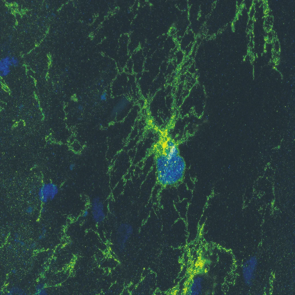
Myelin’s basic structure and function have been understood for decades. It’s a double-thick membrane, similar to a cell membrane, wrapped in tight layers around the axons, or long arms, of our nerves. Open a package of plastic wrap and contemplate the clingy layers snugly circling the cardboard tube if you need a visual here.
Made mostly of fat and protein, myelin works like the insulation on a telephone wire, helping the electrical signal of a nerve impulse travel down the axon more efficiently.
Many of our neurons become myelinated by default. This happens as a normal part of brain development, starting around birth and continuing steadily into middle age. Myelination generally proceeds from the brainstem in infancy and finishes decades later in the prefrontal cortex, our center of complex thinking. (Parents, if your teenagers tell you how dumb you are, feel free to point out that your prefrontal cortex is more myelinated than theirs.)
The idea that there’s more to myelination than this default program — that nerves direct changes in their own myelination as part of learning — was first proposed by the late Stanford Medicine neuroscientist Ben Barres, MD, PhD. As a postdoctoral scholar at University College London in the lab of Martin Raff, MD, Barres showed in 1993 that blocking nerve activity with a toxin had downstream effects on the cells that manufacture myelin. Other clues to the link between myelin and learning came from before-and-after brain scans of people acquiring new skills: The region of the brain where vision and movement are coordinated had more myelin after they learned to juggle, for example.
But uncovering the details of how adaptive myelination works had to await another Stanford Medicine innovation, the 2005 development of optogenetics, a technique in which scientists engineer light-sensitive proteins into specific neurons, creating experimental animals whose nerves can be turned on and off with the flick of a light switch.
Using this method, Monje’s team showed that switching on a subset of the brain’s neurons — motor planning circuitry that, when activated, made the animals walk in circles — could, with repetition, cause specific neurons in that circuit to become more myelinated.
“This discovery was very controversial early on,” Monje said, adding that other neuroscientists were skeptical at first about the new mechanism of learning. (Monje has trained a generation of myelin biologists in her lab and is widely recognized as a pioneer in the field.)
The initial project, published in a 2014 paper in Science, was co-led by Erin Gibson, PhD, then a postdoc in Monje’s lab and now an assistant professor of psychiatry and behavioral sciences. How did the extra myelin get onto the nerves? Gibson’s work showed that the neurons she had experimentally activated sent electrical and chemical signals to nearby neighbors called oligodendrocyte precursor cells. These cells were prompted to divide and mature into another type of brain cell, myelinating oligodendrocytes. These, in turn, added more myelin to nerves involved in the animals’ circling walks, increasing their efficiency.
“Even small changes in myelination can change the speed and synchrony of neural signals,” Monje said. “In the healthy brain, these adaptive changes in myelin ‘tune’ neural circuit dynamics to promote coordinated function.”
When, as a trainee in Monje’s lab, Knowles learned about these discoveries, it got her thinking. The circling mice reminded Knowles of epilepsy, “where you have recurrent, stereotyped patterns of activity, but they’re not helpful patterns,” she said. The discovery set Knowles wondering if worsening seizures could be somehow connected to changes in myelination.
Stopping seizures from worsening
As delicate as the equilibrium of Tony’s life is now, Graglia and his wife, Ashley Evans, worry even more about what will happen as Tony grows. Seizures beget seizures, they know, and people with Tony’s diagnosis usually progress to a more dramatic form, called atonic or “drop” seizures, by their late teens. In these episodes, the person suddenly loses all muscle tone and collapses.
“Atonic drops are horrific: A 200-pound male dropping spontaneously, his head’s getting injured, furniture gets damaged, people get taken out,” Graglia said. “That is the classic SYNGAP1 journey.”
Tony’s parents aren’t easily daunted. Evans works in finance, while Graglia runs an advocacy group for SYNGAP1-related disorders, the SynGAP Research Fund, that the couple founded to fund epilepsy research and support other families with the same condition. They have more resources than many people, meaning not just the money to hire nannies and therapists for Tony but also the time and gumption to ask their neurologists for help getting him onto experimental medications, or to call scientists like Knowles and say, “Hey, you study absence seizures. Did you know our organization gives grants for that?” They are now funding research in Knowles’ lab related to SYNGAP1, including studies of the potential role of maladaptive myelination in the disease and the development of precision therapies.
“Even small changes in myelination can change the speed and synchrony of neural signals. In the healthy brain, these adaptive changes in myelin ‘tune’ neural circuit dynamics to promote coordinated function.”
Michelle Monje, MD, PhD
In the past year, Knowles has also collaborated with another pediatric epilepsy specialist at Stanford Medicine Children’s Health, Christopher Lee-Messer, MD, PhD, who has cared for Tony, to launch a synaptopathy clinic tailored to the needs of patients with SYNGAP1 and a related condition, STXBP1. These rare diseases are known as synaptopathies because they affect genes that play key roles in the function of synapses, the electrical-chemical connections that allow neurons to communicate with their neighbors. “Patients have complex challenges that go well beyond epilepsy, as Tony’s case illustrates, but there have not been any clinics that provide specialized, multidisciplinary care for these kids on the West Coast,” Knowles said.
Even as Tony’s parents do all this work, they ask themselves: How will they protect him from injury?
“He’s 11, he’s already up to here on me” — Graglia gestures to just below his shoulder. “He’s already a big boy, and I’m pushing 50. So there’s this scenario where I’m going to have a 20-year-old dropping on me and I’ll be 60. How am I going to deal with that?”
Interfering with seizures
To understand how seizures change the brain, Knowles, Huguenard, Monje and others studied rats and mice with absence seizures. As in some types of human epilepsy — including Tony’s condition — these animals develop seizures in early life that gradually ramp up.
In a 2022 Nature Neuroscience study, the researchers found that as the animals developed seizures, they had more myelin-forming cells and thicker myelin and more myelinated neurons in brain regions where seizures occur.
The increase in myelin surprised the research team.
“My hypothesis going in was that seizures would somehow interfere with myelination,” Knowles said. “At that point, we thought of myelination as something that happens during normal development and learning, an optimization of brain function. In thinking about disease, I thought we probably would see disruption of that process.”
“It was absolutely unexpected that changes in myelination would have anything to do with seizure progression,” added Huguenard. Before myelin was considered, scientists thought stronger seizures might be explained by changes in nerve-to-nerve connections, via the “original flavor” of neuroplasticity, or flexibility in brain structure, known as synaptic plasticity. “That’s the classic model we think of for development of epilepsy, and this is on a completely different tack.”
Monje was less surprised. “Other studies in my lab around this time were also uncovering maladaptive myelin plasticity in diseases characterized by abnormal levels or patterns of brain activity,” she said.
“Atonic drops are horrific: A 200-pound male dropping spontaneously, his head’s getting injured, furniture gets damaged, people get taken out.”
Mike Graglia
To find out if interrupting the seizure-induced myelination could block the development of seizures, the researchers genetically engineered mice in such a way that they could interrupt the signal from seizing neurons that causes nearby cells to mature into myelin factories. In the genetically altered mice, seizures couldn’t trigger the maturation of more myelin-makers. These mice still had some seizures, but because maladaptive myelination was interrupted, their seizures did not become more frequent over time.
The researchers also used a drug that blocks aspects of the maturation of myelinating cells. The findings were similar to those in genetically engineered mice: Seizures still occurred, but they did not become worse or more frequent.
The class of drugs used in the study, HDAC inhibitors, includes some medications approved by the U.S. Food and Drug Administration. The scientists hope to study whether such drugs could improve outcomes, particularly in children newly diagnosed with severe forms of epilepsy.
“There’s a lot more that needs to be done to explore the molecular mechanisms that link pathological patterns of neuronal activity to maladaptive myelination in severe and refractory epilepsy,” Knowles said.
“This is a beautiful example of translating a scientific insight into an actionable therapeutic tool,” Graglia said. “If Stanford throws its support behind this finding and gets it into humans fast, they can change innumerable lives.”
On a quest for effective therapies
The discoveries highlight the importance for medical science of understanding brain cells that aren’t neurons.
“For most of the history of neuroscience, we’ve focused on neurons and ignored the vast majority of cells in the brain,” Gibson said. For instance, the work of oligodendrocyte precursor cells is only beginning to be explained. Gibson calls them “renaissance cells,” noting they have many jobs beyond maturing into the brain’s myelin factories. And they’re just one example of the many types of glia, non-neuronal brain cells, that we barely understand.
“None of these brain cells live in isolation,” Gibson said. “They live talking to each other and working together, but we’ve always studied them in isolation. I think understanding how these cells communicate — and why, and what the signals are — will open huge avenues of potential therapeutic strategies for neurological disorders.”
Monje adds that “interactions between neurons and glia are essential for normal brain function, and disruption or dysregulation of these powerful interactions can contribute to diseases as diverse as brain fog after chemotherapy, opioid addiction, epilepsy and brain cancers.”
Knowles and her colleagues are exploring how myelination plays into seizures. They recently worked with Jennifer McNab, PhD, associate professor of radiology, to develop a way to use magnetic resonance imaging to measure myelin thickness throughout the brains of research animals with epilepsy. This gives new power to compare myelin changes in different areas of the brain.
“We think changes in the structure of the corpus callosum are unexpectedly playing a role in seizures worsening over time.”
Juliet Knowles, MD, PhD
As they reported in a 2023 paper in Neuroimage, of the two white matter tracts — highways of myelinated neurons — that propagate seizures in their experimental animals, only one tract showed changes in myelination with more seizure activity. It was the corpus callosum, the main link between the brain’s left and right hemispheres. Intriguingly, this was also the main site of change in Gibson’s original research on adaptive myelination.
“We now think the corpus callosum is not just a passive conduit for seizures,” Knowles said. “We think changes in the structure of the corpus callosum are unexpectedly playing a role in seizures worsening over time.”
Knowles’ team is also trying to understand why different types of epilepsy show different patterns of myelin dysfunction. “In temporal lobe epilepsy, which is the most common type, there’s evidence that there is actually myelin loss,” she said. “We don’t know why that disease is different.”
They’re also exploring whether myelin changes might contribute to epilepsy symptoms beyond seizures, including problems with learning, attention or sleep. And they hope to find differences between the biological signals involved in healthy versus maladaptive myelination to use as targets for new treatments.
Still more art than science
Treatments that better match the biology of different types of epilepsy are urgently needed to help doctors match patients to effective medications with less guesswork. Graglia still remembers his first experience with this awkward process: Tony was hospitalized at Packard Children’s, and one neurologist treating him suggested one common anti-seizure drug, ethosuximide, while another neurologist thought it would be better to start with a different drug, lamotrigine.
“I’ll never forget, I’m sitting next to a sleeping baby, he’s sick, he’s 3, in a Stanford hospital, and the first doctor comes back and she’s reviewing the scientific literature and says, ‘Look, here’s a paper saying ethosuximide.’ And the second doctor happened to be there, and he’s like ‘Look at this, saying lamotrigine.’
And I’m sitting on the bed, holding my son’s hand, looking at their badges: they’re both MD/PhDs. That was a major moment for me, realizing it is more art than science.”
Knowles’ team continues to build the science. While their 2022 study in rodents showed that interrupting maladaptive myelination is possible, the practical challenges of making this feasible for human patients are significant. To tackle these challenges, the team is looking for subtle differences between healthy and maladaptive myelination, with the goal of finding features specific to the “bad” version of the process, which could act as drug targets that could stop disease without inflicting harm, Knowles said.
“Many forms of learning are really dependent on this mechanism — so you’d have to develop a therapy that might hit only on pathways in the brain that are specifically involved in seizures, or only at a certain time in development,” Huguenard added. “It could be tricky.”
As researchers face these challenges, it’s worth stepping back to appreciate the beauty and mystery of our wonderfully flexible brains, about which we have so much to learn.
After all, even if it’s been years since you’ve been on a bicycle — even if you’ve grown 6 inches or lost 50 pounds, putting you in a vastly different body than you had last time you rode — you’ll probably be able to swing your leg over the frame and pedal off without a wobble.
Muscle memory, people say. But the memory isn’t in your muscles. It’s in your myelin.
