
Take that
How you clobber the flu infection: A brief tour of the immune system
Let’s say you’ve got the flu. You feel horrible — and you’ve felt that way for days. Hard as it might be to believe, though, chances are excellent that after about a week of this misery, you’ll be feeling better, and within two weeks the symptoms will have all vanished. Here’s how your immune system quashes the virus that set your misery in motion.
Infection
The influenza virus enters through your eyes, nose or mouth and invades the cells lining your airway — the passage from your nose and throat and into your lungs. Its source was probably saliva droplets from an infected person. So far, you don’t feel sick.
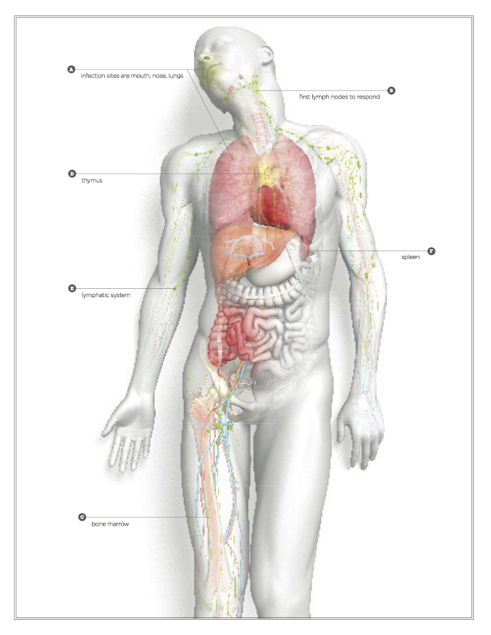
Point A (mouth, nose and lungs on diagram):
(By day 3) The first responders — the innate immune cells — flock to the scene. These blood cells act quickly by either killing infected cells or secreting proteins that recruit other blood cells to the infection site. Among the first to arrive are neutrophils and natural killer cells. Unfortunately, they kill some healthy cells too. This carnage is one reason your throat hurts.
Point B:
(Days 1-4) Macrophages and dendritic cells are also quick getting to the site of infection. While macrophages start killing infected cells, dendritic cells carry intelligence from the infection site to the nearest lymph node. The intelligence is in the form of bits of viral proteins displayed on the dendritic cell’s outer surface.
(Days 4-6) Inside the lymph node, T cells that recognize the viral bits presented by the dendritic cells become activated: They start multiplying and making proteins to help fight the infection. The dendritic cells also release chemicals that trigger some T cells to become T-helper cells, which “give help” by activating other types of immune cells. Other kinds of T cells, known as cytotoxic T cells, become activated to attack infected cells harboring the virus.
(By day 6 or 7) Prepped to attack, T cells travel from the lymph nodes through the blood vessels to the infection site. Other T cells remaining in the lymph nodes help B cells make antibodies with heightened affinity to the viral proteins. Antibodies are specialized proteins that circulate through the blood, stick to pathogens and trigger macrophages to engulf and destroy them.
(Days 7-10) Some B cells develop into plasmablasts. These enter the blood stream and release their antibodies, which attach to the viruses and neutralize their ability to invade cells. Macrophages make a meal of the viruses.
Point C:
(Day 11) It’s likely the infection is now controlled. The immune response winds down. The cells that were activated start dying, though a small pool of memory cells linger, circulating in the blood or residing in the bone marrow.
A brief tour of the immune system
The most common entry sites for infectious agents are through breaks in the skin or through mucosal tissues, including the nose, lungs, mouth and gut.
Point D:
The immune cells are various types of blood cells, all of which originate in the bone marrow. (Though before birth, for a short time blood cells originate in the liver.) Some blood cells migrate to the thymus, where they receive signals that help them become the various types of T cells.
Point E:
The lymphatic system is a network of vessels that connects all lymph nodes in the body. It drains fluid from tissues and organs, and connects to the circulatory system. As a result, the lymphatic system is able to collect antigens from infectious agents (and toxins) and deliver them directly to the lymph nodes for uptake by dendritic cells and presentation to T cells.
Point F:
The spleen cleans the blood of damaged immune cells and microbes tagged for destruction by antibodies. It also contains a full repertoire of immune cells ready to go to work. As such, if your first exposure to a pathogen is through the blood, the spleen will be the main site for immune response.
