
Side by side by side
Saving the Luevanos triplets
When Lily Estrada was six weeks pregnant, she and her husband found out she was carrying triplets. “We were just shocked,” Estrada recalls. The Salinas, Calif., couple, who had three teenagers at home, had been excited about the idea of a new baby. Triplets, though? “My husband almost fainted,” Estrada says.
But the bigger shock was yet to come. As the pregnancy progressed, Estrada’s doctors discovered that her triplets, identical boys, shared a single placenta with a serious defect. During pregnancy, the blood-vessel-rich placenta connects fetuses to their mother and serves as an essential conduit for nutrients and waste disposal. But this placenta was slowly killing Estrada’s three boys.
“It was very bad,” says Yair Blumenfeld, MD, assistant professor of obstetrics and gynecology at the Stanford School of Medicine, who led a team of maternal-fetal medicine experts that cared for Estrada. “Her trajectory meant there was a very high chance she would lose all three babies.”
Early on, the team explained the gravity of the situation to Estrada, who was 35 at the time of the diagnosis in the fall of 2013 and who works in patient registration at Natividad Medical Center in Salinas, and her husband, Guillermo Luevanos, then 36 and an agricultural worker in the Salinas Valley. Estrada’s placenta had several blood vessels connecting the three babies directly to each other, and blood was shared unequally through these connections, a condition called twin-to-twin transfusion syndrome. Researchers don’t know why the disease develops in some cases but not others, though they are trying to figure it out.
Not only was Estrada’s entire pregnancy at risk, at the time no one at Stanford could perform the surgery that might alleviate the problem. But the team could offer something new: Lucile Packard Children’s Hospital Stanford had just entered into a clinical and scientific collaboration on fetal treatment with Texas Children’s Hospital and Baylor College of Medicine, in Houston, which did offer the surgery she needed. Estrada could become their first shared patient.
The collaboration is part of a widespread effort to refine fetal surgery. Excitement about these unusual surgeries rose in the 1980s and ’90s as surgeons attempted to fix many different congenital defects during pregnancy, but the buzz fizzled when the risks became apparent. Cutting into a pregnant uterus often triggers preterm labor, putting not one but two patients — mom and baby — at risk of complications that can easily outweigh benefits of the surgery. Today, surgeons have narrowed the range of conditions for fetal surgery to those with the best chance of averting death or long-term disability for the future baby, and they are developing new tools and techniques to improve safety for both patients.
One big shift: For most diagnoses, surgeons now favor laparoscopic procedures that use tiny incisions and instruments instead of the large, risky, open incisions through the uterus that were once used to access the fetus.
“Over time, people realized aggressive interventions may improve neonatal survival but have very high risk to the mom and the integrity of the uterus,” Blumenfeld says, adding that early techniques left mothers at risk of bleeding and uterine rupture as well as premature delivery.
“Our two teams, at Stanford and Texas, have a very similar perspective on the future of fetal intervention: It is as minimally invasive as possible,” says maternal-fetal medicine expert Yasser El-Sayed, MD, professor and director of Maternal-Fetal Medicine and Obstetrics at Stanford University, and obstetrician-in-chief at Lucile Packard Children’s Hospital Stanford.
Before surgery
Estrada was 17 weeks pregnant when it became clear that something was wrong. On a routine ultrasound, her doctor in Salinas saw too much amniotic fluid. Estrada had some fluid removed, but the problem recurred, prompting her referral to Stanford.
The Stanford team soon realized Estrada’s pregnancy was a perfect storm of rarity. Triplets who share a single placenta occur in one to two births per 100,000, and just a small fraction of these have twin-to-twin transfusion.
“With a shared placenta, there are always going to be some vascular connections between the babies,” Blumenfeld says. Although most of the blood from each baby’s umbilical cord travels deep into the placenta to exchange nutrients and wastes with mom’s circulation, some blood flows into surface blood vessels that connect the babies directly to each other. In 10-15 percent of shared placentas, these connections lead to the unequal blood flow that characterizes twin-to-twin transfusion. (The condition is so named because most cases affect identical-twin pregnancies, which are much more common than triplets.)
Shared placentas with abundant artery-to-artery connections between fetuses seem not to develop problems, researchers have noted, whereas those with predominantly artery-to-vein and vein-to-vein connections are primed for uneven blood flow.
“The baby pumping blood has to work very hard and can get very sick, and the recipient baby, who is getting a lot of extra fluid, can go into heart failure,” Blumenfeld says. In the 1980s, before surgical treatment was developed, the medical literature reported 95 percent mortality of fetuses like Estrada’s that were affected by a severe fluid imbalance before 24 weeks of pregnancy. Surgery on the placenta has improved survival substantially, but many fetuses still die before birth. And the high mortality is especially sad because, in most cases, the fetuses are otherwise normal.
In Estrada’s case, one fetus was donating blood through the placenta to the second, who was passing it through other shared blood vessels to the third, and sickest, fetus. Although they shared a placenta, each brother had his own amniotic sac, the “bag of waters” in which a fetus develops. Amniotic fluid, the cushioning liquid in the womb, was accumulating in the third fetus’s amniotic sac as his kidneys produced extra urine to lighten the fluid load from the excess blood, while the fluid in the sacs of the donor fetuses dwindled away to almost nothing.
Extra fluid was changing the structure and function of the recipient’s heart, putting him at risk for heart failure before birth. Not only was his health in danger; if he died in utero, his brothers could suffer permanent neurological injury. (No one is sure why this happens — perhaps a sudden change in blood pressure or rush of cytokines at the time of the death is responsible — but experts agree that the brain injury to surviving fetuses seems to occur instantaneously.)
‘The baby pumping blood has to work very hard and can get very sick, and the recipient baby, who is getting a lot of extra fluid, can go into heart failure.’
Making a decision
The Stanford team gave Estrada and Luevanos several options: End the whole pregnancy, do nothing and face the hazards of the condition, terminate one fetus’s life in the hopes of improving the chances of the other two, or have Estrada travel to Texas for surgical treatment that uses a laser to seal off problematic blood vessels in the placenta.
The medical team faced a delicate balance of trying to convey the potential benefits and risks of each option without raising falsely high hopes. They also wanted to give the couple enough information to make an informed decision but enough room to feel like they were in the driver’s seat, even if the options were potentially heartbreaking.
“It’s a very difficult situation for a family to be in,” says neonatologist Susan Hintz, MD, medical director of Fetal and Pregnancy Health Services at Lucile Packard Children’s Hospital Stanford and one of the leaders of the Stanford-Texas Children’s Collaboration. “We do not want to be paternalistic about conveying the options, especially in a very challenging and rare case such as this one. We’re working with a family to understand what their goals are, and we want to be honest about our estimates of the risks and the limits of what we can predict with certainty about the outcomes.”
In Estrada’s case, no one was sure how well the surgery might work because so few triplets are treated for twin-to-twin transfusion.
“We were saddened and sort of confused,” Estrada says, recalling the first reactions that she and her husband had to the news. “It was: We could wait and see what happened, but the likelihood was that we were going to have no baby, or we could terminate one and see what happened with the other two, or take the risk, go to Houston, have the surgery and hope it worked for all three. But they didn’t guarantee anything.”
One piece of background that helped inform the couple’s decision was the fact that when the surgery worked, research had shown it helped moms stay pregnant about four weeks longer, allowing their babies more time to develop before birth. (Because the uterus gets so crowded, twins and other multiples are almost always born early, but a less premature delivery makes a huge difference for the babies’ health.) Sealing the connecting blood vessels also seemed to protect surviving fetuses in the event that one died. “We’re separating, or attempting to separate, their fates,” Blumenfeld says.
After a lot of counseling and discussion with the Stanford team, “we decided to go for it and do surgery,” Estrada says.
Once they had made the choice, they had no second thoughts. “My husband was a little bit stronger,” Estrada recalls. “He just wanted me to go for it, and see what happened.”
Interacting with Estrada and her family had left Blumenfeld, like the rest of the Stanford team, anxious to do the best he could for her.
“She’s an absolute gem to take care of,” he says.
In the operating room
Estrada’s flight to Houston, when she was 21 weeks pregnant, was difficult in itself. She was traveling alone; Luevanos had to stay home with their older children, while Blumenfeld, who was coming to observe and help manage the patient, was following on a later flight. The accumulated amniotic fluid added challenges at every step. “It was scary,” Estrada recalls. She was very uncomfortable sitting still, had trouble drawing a deep breath and had enough difficulty walking that she needed a wheelchair to navigate the airports. When the four-hour flight to Houston took off, her uterus swelled even more, putting her whole belly under extreme pressure. “It was a mission just to get from one place to another,” she says.
When she arrived at Texas Children’s on the evening of Dec. 14, 2013, she began spotting — just a little vaginal bleeding, but it was still worrisome. Although the bleeding had stopped by the next morning, echocardiograms showed the babies were in distress, and the operation was moved up a day. Blumenfeld came directly from the airport to Texas Children’s, arriving at 3:30 p.m. on Dec. 15, just as Estrada was being wheeled into surgery.
Estrada’s surgeon, Michael Belfort, MD, PhD, obstetrician- and gynecologist–in-chief at Texas Children’s Hospital, had two goals: to remove excess amniotic fluid and, more important, to seal every abnormal blood vessel connecting the babies that he could find. He aimed to avoid shutting down any vessels that were not involved in the transfusion syndrome so that each baby would get the maximum benefit of his piece of the placenta.
But although Belfort would try to seal off every problematic blood vessel, in a uterus crowded with three babies, he knew he might have difficulty finding them all. And it would take about a week of monitoring the babies’ fluid levels to see if the procedure had helped.
“We do the best we can,” says Belfort, chair of Baylor’s Department of Obstetrics and Gynecology, who has performed more than 200 surgeries for twin-to-twin transfusion since he learned the procedure in 2007. Even with his years of experience, though, Estrada’s was only the second case of triplets he had treated. “If we’re able to identify all of the abnormal blood vessels, we are definitely able to help. If we can’t identify them all, we maybe get partial resolution.”
In the operating room, Belfort made a 10-millimeter incision and inserted the surgical tool, which contained both a tiny camera and a laser, into the amniotic sac of the triplet who was receiving blood flow from his brothers. The view on the monitor in the operating room — a round window into the babies’ prenatal world — showed a jumble of pale limbs, glimpses of umbilical cord, a flash of a tiny ear, and, near the top of the field of view, the network of blood vessels on the surface of the placenta. The recording of the surgery illuminates a prenatal world most people never see. It’s entrancing, a little scary and hauntingly beautiful. Watching it, one can’t avoid imagining the hopes of everyone in the operating room: that the surgery would work, that the babies would keep growing, that they would be born safely, that Estrada and Luevanos would get to hold their three infant sons and kiss them and tell them everything was going to be OK.
To begin, Belfort looked for the fetuses who were pumping blood onward, known as “donors.” He could identify them by the fact that they were in nearly empty amniotic sacs and appeared trapped behind the very full amniotic sac of the triplet receiving the most fluid.
Once he had identified the donors, Belfort began visually tracing blood vessels that ran laterally across the surface of their placenta to the recipient. Each vessel got a zap of green laser light to seal it. He aimed the laser at the point where the vessels met between two fetuses, or at a spot as close to that as he could reach that allowed him to shut down the connection. In total, he sealed seven blood vessels, a typical number for this type of surgery, then made some shallow passes with the laser to catch other small blood vessels on the placenta’s surface that he might have missed. He also removed about a liter of amniotic fluid from the recipient’s amniotic sac. The entire procedure took about an hour.
Better tools
Blumenfeld’s trip to Texas to observe Estrada’s procedure was the first step in building the surgical skills of the Stanford team, a key aspect of the Stanford-Texas collaboration. Since then, both Blumenfeld and Stanford pediatric general surgeon Karl Sylvester, MD, obtained Texas medical licenses to enable them to get hands-on training in Houston, and Lucile Packard Children’s Hospital Stanford ordered new surgical equipment for fetal interventions. The team conducted their first surgery for twin-to-twin transfusion syndrome at the hospital in January.
“In fetal surgery, the surgeons need better hands and better eyes,” says Christopher Contag, PhD, a Stanford professor of pediatrics and of microbiology and immunology who builds medical-imaging and visualization tools for surgeries. With the shift to minimally invasive techniques for fetal procedures, surgeons are limited by the cameras and laparoscopic instruments already on the market. Better “hands” would be a big help in one common fetal surgery: repairing spina bifida, a congenital defect in which portions of the spinal cord are exposed to the exterior of the body. Ideally, surgeons would like to have dextrous laparoscopic tools to hold the needle they use to repair spina bifida. But now, their tools have so little articulation that they’re forced to make stitches by passing the needle back and forth between two surgeons.
For twin-to-twin transfusion surgery, the “hand”— the laser used to seal blood vessels — is already extremely effective. But surgeons want better eyes: “Doctors need to see the blood vessels, and need to have them stand out against background,” Contag says.
Right now, the light source used to look for problematic placental blood vessels is ordinary white light. Under that illumination, it’s tricky to pick out the blood vessels and impossible to check whether a cauterized vessel has been sealed.
But blood’s intrinsic spectral properties will probably make it possible to solve these problems. For finding blood vessels, “we can illuminate the tissue at one wavelength and collect light at another,” Contag says. “It makes the blood vessels jump out at you.” (His team has already incorporated this technique into tools they’ve developed for gastrointestinal endoscopy.) To check the seal on cauterized vessels, surgeons could use photoacoustics to listen for blood flow: You shine laser light at the blood vessels, which absorb some energy, heat, expand and reflect back patterns of ultrasound waves that can be heard with ultrasound transducers and transformed into 3D images. “You can very clearly see if you were successful,” Contag says.
Happy Birthday
After the surgery, Estrada stayed a week in Houston for monitoring. Since surgical tools that verify cauterization of blood vessels are still in the future, the best way to confirm the success of the surgery was to watch what happened to the babies. “The proof is in the pudding,” Texas Children’s Belfort says.
The fetus that had appeared least affected by fluid imbalance before the surgery developed worrying swelling in his skin and umbilical cord in the first day after the procedure. At first, the doctors weren’t sure if he would get better or worse.
‘If we’re able to identify all of the abnormal blood vessels, we are definitely able to help. If we can’t identify them all, we maybe get partial resolution.’
Waiting to find out what would happen was taxing for Estrada. Separation from her family and worry about her triplets left her feeling lonely and depressed.
But good news was coming. “Within days, we saw improvement,” Belfort says. The worrying swelling abated, and ultrasound scans showed all three fetuses had reassuringly normal-sized bladders, a sign that their bodies were not fighting the fluid imbalance that affected them before surgery.
With less amniotic fluid, the trip home was much easier for Estrada. After returning to California, she stayed in an apartment near the Stanford campus with her husband; the medical team wanted her close to the hospital for delivery. Triplet pregnancies last an average of 32 weeks, eight weeks short of the 40-week gestation period that is normal for single babies. To reduce the effects of prematurity, the doctors wanted to get Estrada as close to that goal as possible, but weren’t sure how long her pregnancy would last. “Every day we get now is a gold mine,” Blumenfeld said at the time.
On Jan. 30, 2014, when Estrada was 28 weeks pregnant, she went into labor. Her due date was still 12 weeks away, but the babies had a strong chance of surviving the effects of prematurity. At the hospital, Blumenfeld determined that one fetus — the baby who had been receiving fluid before surgery — had a falling heart rate, so an emergency C-section was performed.
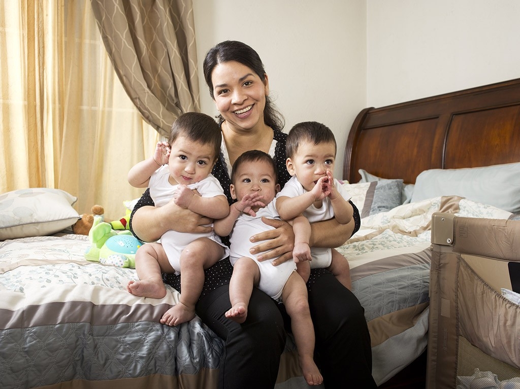
Baby Pedro was born first, weighing 2 lbs. 9 oz. Then came his brother William, at 2 lbs. 6 oz., and finally little Ayden, the recipient of the fluid, at 1 lb. 9 oz.
“I was emotional,” Estrada says. She couldn’t see the new arrivals in the delivery room, but she heard their three first cries. Later, when she was wheeled on a gurney to meet them in the neonatal intensive care unit, “I was scared for them because they were so tiny,” she recalls.
Although the babies all had complications of their premature arrival, under the ministrations of the neonatology team they gradually matured and gained strength. Ayden, the baby who had been most severely affected by twin-to-twin transfusion, also had the most difficulty after birth, but was ultimately able to go home to Salinas in April, just a week after his brothers. He still gets checkups with Stanford Children’s Health specialists every six months to monitor potential complications of premature birth, including possible problems with his lungs and his vision, but his doctors are encouraged by his progress.
Today, though Estrada and Luevanos feel the exhaustion one would expect of parents of young triplets, they’re also delighted to be watching all three of their little ones grow. Pedro and William are crawling, playing peek-a-boo and trying to walk; and Ayden, though a bit behind his brothers developmentally, is rolling everywhere and picks things up off the floor. “They’re really happy babies,” Estrada says. “They’re doing well.”
The Stanford team, meanwhile, is looking forward to being able to help other patients with twin-to-twin transfusion. “This is one of the greater success stories of fetal intervention: It’s minimally invasive, the alternative of no treatment is horrific, and the surgery gives you a chance of taking home two babies if you have twins or three babies if you have triplets,” Blumenfeld says. “Before, there wasn’t this opportunity.”
