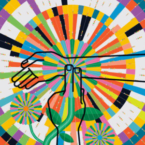
Listen up
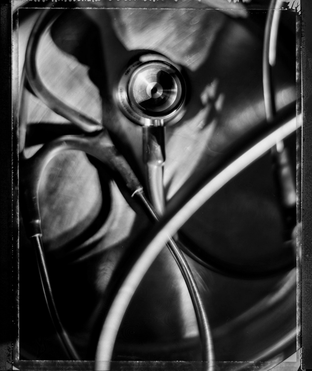
The stethoscope at 200
John Kugler, MD, gently places the stethoscope on the upper chest of the elderly, white-haired woman who is sleeping peacefully while propped up in bed, her head cocked to one side as a dialysis machine clicks away in the background. He spends a few seconds listening to her heart and lungs, which have a crackling sound — a sign of possible fluid in the lungs or another respiratory problem. Her breath sounds are faint.He then reaches into his black nylon bag for his other ever-present medical tool, a portable ultrasound machine the size of a smartphone. He gingerly lifts the patient’s light-green gown so as not to disturb the dialysis wires, and after massaging some gel on her belly, he applies the ultrasound probe to view her lungs on the small screen.
“She still has a pretty big effusion there,” he tells the medical student, pointing to a spot on the screen where he sees a collection of fluid. “It’s going to take some time for that fluid to be absorbed.”
The ultrasound can also reveal some signs of infection or inflammation, things he can’t discern by listening with the stethoscope.
“So we learn more by using the ultrasound. It’s not a magic wand, but it’s useful,” says Kugler, a clinical assistant professor of medicine at Stanford.
The stethoscope — that time-honored symbol of the medical profession — is still the first line of diagnostic inquiry for most clinicians, but it is losing ground to imaging technologies that can yield more precise and expansive information about a patient’s condition. And as its stature fades, so is the fine art of listening to the inner workings of the body.
Does the 200-year-old instrument have a place in medicine’s future? It depends on whom you ask.
A 200-year history
Since the time of Hippocrates, physicians have relied on sound to diagnose physical ailments: To listen to the heart beat they simply put their ear to the patient’s chest. The modern-day stethoscope first came to life in 1816 when René Laennec, a reportedly shy French physician, encountered a plump young woman with an apparently diseased heart. As the story goes, he was too embarrassed to lay his ear to her ample chest, so, inspired by seeing children in a Paris park scratch at one end of a piece of wood while listening at the other, he rolled up a piece of paper into a tube. When he placed the crude device against the woman’s chest, he was delighted to hear distinct heart sounds, better than any he had heard before. He soon switched to wood, designing his first, foot-long prototype in pine, with a funnel-shaped piece at the end for listening. By examining patients on autopsy, he began to correlate the sounds he heard with specific problems in the heart and lungs.
Laennec christened the device the stethoscope — Greek for looking (scope) into the chest (steth). The initial model underwent various changes, including the introduction of other materials, such as ivory, silver and brass, but the biggest advancement occurred in the 1850s with the introduction of the binaural stethoscope, with flexible rubber tubes used for listening with both ears, which dramatically reduced external noise and improved quality of sound. That basic design is not all that different from the stethoscopes doctors dangle around their necks today, says pediatric cardiologist Daniel Bernstein, MD.
The refinement of the device ushered in the golden era of auscultation, the listening art that became an essential part of the physical exam. The stethoscope remains quite simple in design, consisting of the chestpiece, which is placed against the patient’s chest, and two tubes that carry the sound from the chestpiece to each ear. One side of the chestpiece is the diaphragm, a plastic disc, while the other side is a bell, a hollow cup. When either side is placed on the chest, the body’s sounds cause it to vibrate, creating acoustic pressure waves that are carried through the tubes to the listener’s ears. The bell picks up low-frequency sounds, while the diaphragm picks up sounds of a higher frequency.
The stethoscope came to be commonly used for detecting heart and lung problems — like the whoosh of a heart valve that is not closing properly or the crackling sound of pneumonia. It also became valuable for detecting abnormalities in the digestive and vascular systems, such as the gurgling that may accompany an obstructed bowel.
‘When I was a student, you walked around the wards with your attending physician and everybody listened. I think those skills are at risk of being lost.’
Longtime practitioners like Bernstein recall the days of the master diagnosticians in his field of pediatric cardiology, who could discern multiple aspects of the quality of a murmur and use it to identify not only a specific cardiac anomaly but the severity of that anomaly simply by listening to the heart, refined skills that he fears are fast disappearing.
“When I was a student, you walked around the wards with your attending physician and everybody listened,” says Bernstein, the Alfred Woodley Salter and Mabel Smith Salter Endowed Professor in Pediatrics. “You had 10 white coats in a room, and everybody took their turn to listen to the patient. That way everyone learned. Now there are 10 clinicians with computers on wheels, and few examine the patient. So I think those skills are at risk of being lost.”
Concerned about the decline of basic bedside skills, Abraham Verghese, MD, a professor of medicine at Stanford, created the Stanford 25 — a set of 25 essential exam skills — nearly 10 years ago to help reinforce the practice and the importance of the physical exam in diagnosis, including auscultation. In addition to regular sessions for trainees, he brings clinicians to Stanford from around the country as part of a movement to keep alive the culture of bedside medicine.
“I would emphasize that there is a ritual to the doctor-patient encounter. Patients undress and allow you to touch them, which in any other context would be viewed as an assault. So they give you this great privilege,” Verghese says. “There is a craft to this, and if you don’t do it with skill, patients pick up on that.”
He says the stethoscope is a key element of this ritual and can provide a “piece of the puzzle” for diagnostic purposes. “The stethoscope allows me to very quickly discern some information, and the ultrasound allows me to refine that. So they are additive. What’s important is that you use these instruments and use the exam well.”
The technology challenge
The steady erosion of physical exam skills began in the 1970s with the advent of new imaging technologies, such as MRI, CT and particularly ultrasound, a painless, radiation-free tool that uses sound waves to create a moving visual of the internal organs. Clinicians now could directly view the anatomy beneath the surface with great precision. While the early ultrasound machines were bulky devices that had to be wheeled into a room, they have progressed to handheld versions with greatly improved visual clarity, produced at increasingly reduced cost.
Many physicians now routinely carry these pocket-sized devices on their rounds, while larger, portable ultrasounds, resembling a computer laptop, have become standard fare in hospital intensive care units and emergency rooms.
In the past, while clinicians might have spent 15 minutes using a stethoscope to discern the quality of a heart murmur, they may do a quick listen, then order an echocardiogram, an ultrasound of the heart, says pulmonologist and critical care specialist Ann Weinacker, MD.
“You don’t have to spend 15 minutes or so trying to figure out what you think you hear and putting patients through various maneuvers,” says Weinacker, a professor of medicine. “You can just put an ultrasound on their chest and find out. And there are measurements you can get with an ultrasound that you can’t get with a physical exam — or not very easily.”
For instance, she says nowadays it’s possible to do an ultrasound of the lungs, something not commonly practiced just five years ago. Among other things, the test can show the severity of a collapsed lung — something that may not always be discernible by listening alone. It can also show the extent of fluid around the internal organs — a possible sign of heart failure or other problem — and be used day after day to measure fluid changes without exposing patients to ionizing radiation, as an X-ray or CT scan would.
“The truth is if you use technology well, you can get a lot more information,” says Weinacker, who routinely uses it in the intensive care unit to assess a patient’s status.
Jagat Narula, MD, PhD, professor and chair of cardiovascular medicine at Icahn School of Medicine at Mount Sinai, is among those who believe the stethoscope has become a “vintage accoutrement,” rightfully supplanted by swiftly advancing imaging technology.
“It has outlived its time,” says Narula. “Now I can clearly look into the chest, and not only the chest, the whole body. … You have a much superior thing in your hand,” he says of the ultrasound. “Why would you not use it? The stethoscope is obsolete. We should write an obituary for it.”
Just listen
Yet the stethoscope continues to inspire devotees in part because it sometimes works better than anything else, at least in their hands — and ears. Certain problems that would not be detected by ultrasound can be discerned by listening, such as the wheezing of a patient with asthma or with chronic obstructive pulmonary disease, Kugler says. Listening also may point the clinician down a path to diagnosis, providing guidance, for instance, on where to direct the ultrasound probe to confirm a suspected problem.
Bernstein says clinicians can overlook serious conditions in young patients if they fail to do a thorough clinical exam, including listening carefully with a stethoscope. For example, he has encountered children with coarctation, or a narrowing of the aorta, a congenital problem that can stress the heart and compromise the cardiovascular system. “I’ve seen 18-year-olds with coarctation where the diagnosis has been overlooked because nobody did a good physical exam,” he says.
And there are instances, he says, where the results of an echocardiogram may be inconclusive or conflict with something on the physical exam. Once, while examining a patient in a pediatric cardiology outreach clinic in San Luis Obispo, he noted a whooshing sound over the left side of the patient’s chest. The initial echocardiogram did not show any abnormality, but with this discrepancy between the clinical findings and the ultrasound, Bernstein and the ultrasound technician persisted, and were able to find a tiny but potentially life-threatening tear in the wall of the aorta. The patient was transported to Lucile Packard Children’s Hospital Stanford, where his aorta was successfully repaired. “Had we relied only on the initial ultrasound, this could have been a disaster,” Bernstein says.
For the basic care of newborns, the stethoscope is essential, says William Benitz, MD, the Philip Sunshine M.D. Professor of Neonatology. It’s needed for checking a baby’s heart rate or listening to the heart and lungs for possible signs of a major anomaly, such as a diaphragmatic hernia, an abnormal opening of the diaphragm.
“In a matter of a few minutes, you can move from not knowing very much about a baby that is not behaving very well to having a specific diagnosis, and it’s all based on a stethoscope,” Benitz says. “So I don’t think we’re on the verge of replacing physicians with machines just yet. But I do worry we’re not training our young people to trust their exam skills and ask the right questions and trust their intuition. They have so much more to learn than we did 30 years ago. We just have to strike some kind of balance.”
Skills set
Some younger physicians acknowledge they don’t rely on the stethoscope the way their older counterparts do.
“A lot of people know there will be an imaging or ultrasound exam that they are going to do anyway and because we are less likely to make critical decisions without having the information from imaging, inevitably clinical skills will not be as robust as they were years ago, when that was all you had,” says Andrew Chang, MD, co-chief resident in internal medicine at Stanford. Nonetheless, trainees and younger practitioners still value the stethoscope as a diagnostic tool, he says.
“Even if individuals say young doctors aren’t as attuned to the sounds that come through their stethoscopes — and I do think there is less of an emphasis on this and we aren’t as well-trained in this — I still feel it’s something we heavily rely on,” says Andre Kumar, MD, co-chief resident in internal medicine, who says he would feel “naked” without his stethoscope.
Both he and Chang are interested in global health and have found themselves in places like Uganda, Bangladesh and Nepal, where resources were limited, ultrasound a luxury and stethoscopes absolutely essential for diagnosis. These situations exposed the importance of basic skills, they say.
“There were so many times when I felt woefully unprepared to diagnose what was wrong with my patients, sometimes as a direct consequence of my physical-exam skills,” Kumar says.
But one needn’t stray far from Palo Alto to encounter situations where a low-tech approach to diagnosis is extremely valuable, as in a middle-of-the-night emergency when imaging isn’t available, or in a clinic where the cost of a scan may be out of reach for patients, says Lars Osterberg, MD, MPH, associate professor of medicine.
“A great example is the Cardinal Free Clinics. For a while, we didn’t have chest X-rays, and I would diagnose pneumonia without X-rays, which could cost the patient $100,” Osterberg says. “We forget that we are in a privileged society that has access to all these things. That’s not always the case. Just down the street or across the highway people can’t afford certain things. If we did an ultrasound or an X-ray on everyone, it would add up.”
The cost of a cardiac ultrasound varies widely, with Medicare reimbursing between $153 and $698, depending on the type of test, according to published data. And while the cost of ultrasound machines has been steadily declining in recent years, a stand-alone or portable device may cost as much as $100,000. Clinicians typically don’t charge patients when they use a handheld ultrasound during exam, but the device itself can cost at least $8,500. By contrast, “a really decent $50 stethoscope can go a long way,” Osterberg says.
Looking to the future
Most important, some clinicians argue, is the role of the stethoscope as an enduring symbol of the physician and as the center of the ritual encounter between physician and patient.
The stethoscope “puts you close to the patient,” says cardiologist Eddie Atwood, MD, a professor of medicine who has been practicing for more than 40 years. “You’re leaning in. You’re touching the patient. But psychologically, and more important to me, it makes you think about your patient. You spend more time with the patient. By virtue of putting this on, you are right in the patient’s space and thinking about him while you’re working. … That may never go away. This is an element that maybe shouldn’t go away.”
To recognize the stethoscope’s signature role in medical practice, medical schools like Stanford pay homage to it every year in a ceremony in which each incoming medical student receives his or her first listening device.
“Maybe in the future, instead of the stethoscope ceremony, you will get an ultrasound probe,” says S.V. Mahadevan, MD, associate professor and chair of emergency medicine. “We may see the evolution as that being a symbol of our profession.”
But for now, the stethoscope retains its stature. “This stethoscope represents the physician-patient connection,” Lloyd Minor, MD, dean of the School of Medicine, told the 93 incoming medical students during the Aug. 26 ceremony. “When you wear this, you commit to the physician-patient relationship above everything else.”
