


From a car jack to precision cancer care
Three Stanford medicine inventions that made radiotherapy safer and more precise


It was early 1956 when Henry Kaplan, MD, the first chair of the Stanford University School of Medicine’s department of radiology, ran an unusual errand.
After years of hard work, Kaplan and his colleagues were almost ready to fire up their new invention, the country’s first medical linear accelerator. The machine transformed a beam of speeding electrons into a stream of high-energy X-rays capable of penetrating deeply into the body to irradiate tumors, offering the hope of vastly improved cancer treatment.
But as they prepared to treat the first patient, the doctors realized they needed one more piece of equipment. So, Kaplan set out from the teaching hospital — located, like the medical school, in San Francisco at that time — to visit a nearby auto garage.
“I will never forget the puzzled look on the face of the garage owner down on Fillmore Street when I asked him to borrow a heavy-duty automobile jack,” Kaplan said in an interview later quoted in The World of Stanford Radiology, a history book published in 2006, after his death. “I explained that it was to carry a large block of lead with a pinhole in it.”
Key takeaways
- Radiation is the most widely used cancer therapy. About two-thirds of cancer patients in the U.S. receive it.
- Since the mid-20th century, Stanford Medicine experts have built a tradition of innovation in radiation therapy, helping doctors treat more patients and a wider variety of conditions with greater safety and effectiveness.
- The advances help patients of all ages but are especially beneficial for children.
The block of lead — and the pinhole — worked with the new Stanford Medicine linear accelerator technology to deliver radiotherapy as precisely as possible. The physicians needed them because their patient was a baby boy with an eye tumor. To preserve his vision, they wanted to administer a narrow stream of X-rays to miss the lens and cornea of the child’s eye. Hence, the odd errand.
Kaplan’s trip to the local garage was an early step in establishing a tradition of outside-the-box decision-making that many Stanford radiotherapy innovators would follow, to the benefit of generations of patients at Stanford Medicine and around the world.
For decades, Stanford has pushed radiotherapy toward tighter targeting and fewer side effects — starting with the first klystron-powered medical linear accelerator, later with the first image-guided radiosurgery, via CyberKnife, and now a way to broaden access to proton therapy.
Targeted radiotherapy, with fewer side effects
Since the early days of their field — which began with parallel efforts at Stanford and in England to develop the world’s first two medical linear accelerators — radiation oncologists, neurosurgeons and others who rely on radiotherapy have faced the same challenge: how to deliver radiation exactly where it needs to go, with as few off-target effects as possible.
Radiation kills cells by damaging their DNA. It’s good at eliminating malignant cells, especially those buried inside delicate body structures that would be damaged by surgery. It can also be used to destroy other harmful tissues, such as the tangled blood vessels in an arteriovenous malformation, which can rupture and cause strokes if left untreated, or neurons that trigger focal seizures in people with epilepsy.
But radiation also poses risks. If it hits healthy tissue, radiation can cause short-term injury as well as long-term problems such as secondary cancers and organ damage from inflammation and scarring.
Stanford Medicine experts have a long tradition of leading the development of radiotherapy technologies that advance the precision, accessibility and applications of radiotherapy. Here are three of their key inventions:
LA-1: North America’s first medical linear accelerator
Kaplan arrived at Stanford in 1948, drawn in part by the university’s research on particle accelerators. Stanford University engineers and physicists, experts in the nascent fields of microwaves and radar, had already made discoveries used in radar systems that defended Great Britain from World War II air raids.
A key innovation was the invention in 1937 of a microwave power amplifier, the klystron, by brothers Russell and Sigurd Varian working with Stanford University physics professor William Hansen, PhD.
Microwaves could also be used to speed up electrons to very high energy levels. Hansen, working with physics department colleagues including Edward Ginzton, PhD, invented novel designs of particle accelerators called linear accelerators, using klystrons as the source of microwave power, for high-energy physics research.
Kaplan sought out Ginzton to turn linear accelerator technology into a medical device capable of zapping cancer cells. Radiologists were already treating cancer with lower-energy X-rays or by directly administering radioactive substances to tumors (through tubes, hollow needles and other devices).
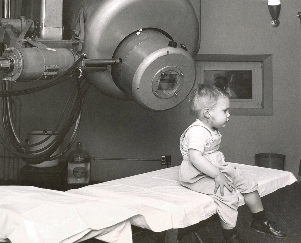
But these methods made it hard to treat tumors deep inside the body and exposed patients to a lot of off-target radiation. Aiming the sped-up electrons from a linear accelerator at a metal target transformed them into high-energy X-rays that could penetrate more deeply into the body.
Kaplan and Ginzton raised funds for the project; supervised construction of equipment in physics labs at the university campus in Palo Alto; and oversaw installation of the machine, dubbed LA-1 (for Linear Accelerator 1), in a concrete bunker built into a San Francisco hillside, adjacent to what was then Stanford’s hospital. (Plans to move the medical school to Palo Alto were underway, so the bunker was designed with a hatch that would allow the machine to be lifted out later.)
Their first patient was a 7-month-old boy who had a genetic form of retinoblastoma that affected both eyes. Before Kaplan’s team met him, surgeons had removed his right eye. The radiologists wanted to do everything possible to spare his remaining vision.
The linear accelerator-based X-ray therapy, precisely shaped by the block of lead pierced with a pinhole, worked: The team irradiated the child’s tumor while avoiding the lens and cornea of his left eye, and he lived for decades after treatment with his vision intact.
LA-1 was moved to the Palo Alto campus in 1959 and used to treat patients until 1972, when it was decommissioned and acquired by the Smithsonian Institution. The most recent medical linear accelerator to be installed at Stanford Medicine Cancer Center, LA-20, is a descendant of the original LA-1 design.
CyberKnife: Image-guided radiotherapy with submillimeter accuracy
As radiotherapy continued to evolve, experts around the world developed better and more specialized ways to deliver radiation. One such innovation, the Gamma Knife, was invented in Sweden in 1967 by Lars Leksell, MD, PhD, to treat diverse brain disorders. The patient’s head is stabilized in a frame, then positioned inside a fixed, dome-shaped array of many cobalt-60 radiation sources.
The physician uses imaging data about the location of the problem to guide beams of gamma radiation from the radioactive cobalt, which converge from multiple angles on the tumor (or other surgical target). The target receives a high radiation dose, but because the beams arrive from many directions, other parts of the head get much lower doses.
In the 1980s, John Adler, MD, then a young neurosurgeon studying with Leksell in Stockholm, was so struck by this concept that he decided to become a medical inventor himself.
“I knew I wanted in on this new thing,” Adler said recently. He is now a Dorothy professor of neurosurgery, emeritus at Stanford Medicine. “I thought it was the coolest thing I ever saw.”
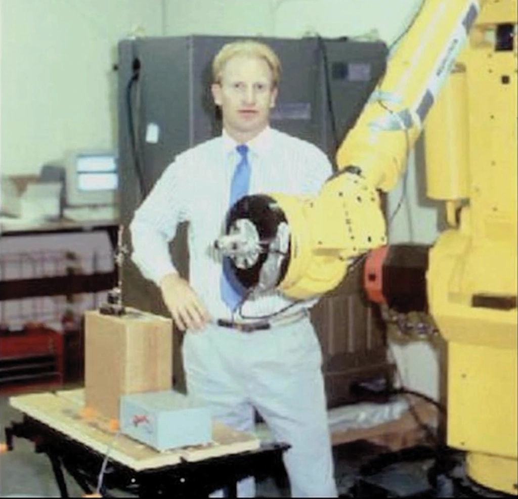
The Gamma Knife reminded him of the way a magnifying glass can focus sunlight. Its relative noninvasiveness soon had him thinking of difficult surgical problems he wanted to use it to tackle. But it had a big drawback: The shape of the Gamma Knife and associated head brace meant you couldn’t treat anything outside a patient’s head.
“I thought, if this concept of convergent beams works so well, might it not work anywhere in the body?” Adler said. “That was my inspiration.” Instead of using several fixed radiation sources to generate convergent beams, he tried moving a single radiation source around the patient.
Adler came to Stanford University in 1987 to put his idea into action. It was the right place for his complex project to come together because there were so many experts here who had elements of the knowledge he needed.
“Perhaps most lucky of all was the proximity to people who knew how to make linear accelerators,” Adler said. He also began working with experts at Stanford University’s School of Engineering on a new approach for noninvasively localizing a target inside a patient’s body, the principles of which would start the field of image-guided radiation therapy.
The new machine — eventually named the CyberKnife — was designed around a new computational method for using simple skull X-rays to precisely aim the radiation beam at a surgical target deep inside a patient. The concept is similar to the way the brain merges images from our two differently positioned eyes to give us accurate depth perception.
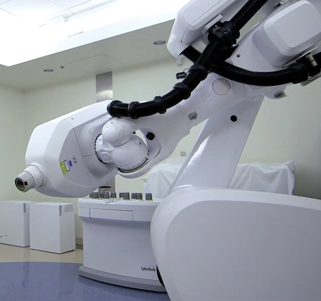
“You acquire X-ray images from two different directions, computationally compare those images and compare them with the CT scan,” Adler said. “Today, it’s almost instantaneous. You can determine where the patient’s head is positioned, and should their head move even slightly, a new and different X-ray projection enables retargeting. It became the first viable means to do precision image-guided tracking within the field of radiation.”
Advancing the CyberKnife from early concepts to a working device took several years, many rounds of fundraising, and resilience to withstand the skepticism of other neurosurgeons and radiation oncologists. (Its inventor jokes that the device was known for a while as “Adler’s folly.”) Stanford Hospital patients were the first to be treated with CyberKnife, in 1994.
Gradually, the machine came to be recognized as a valuable addition to surgeons’ and radiation oncologists’ tool kits. The CyberKnife can be aimed with less than a millimeter of error when delivering radiation, minimizing side effects. It enables noninvasive radiosurgery, which has revolutionized treatment for conditions where conventional surgery would cause a lot of damage to healthy tissue.
The top applications include metastatic tumors and arteriovenous malformations of the brain and spinal cord, as well as cancerous and noncancerous conditions of the head and neck, heart, lungs, prostate and abdomen — such as liver and kidney tumors.
Today, hundreds of CyberKnife machines have been installed in hospitals around the world.
Proton radiotherapy arrives at Stanford Medicine
On April 7, 2026, Stanford Medicine radiation oncologists cut the ribbon on the latest example of their innovative prowess: a first-of-its-kind facility, formally known as the Sridhar B. Seshadri Proton Therapy Suite.
Proton therapy enables oncologists to deliver cancer-killing radiation precisely to a tumor with minimal damage to healthy tissues. Although it was developed in the San Francisco Bay Area in the 1950s, this therapy has been hard for Northern California patients to obtain. The football-field-sized footprint of traditional equipment and its high cost prevent most hospitals from offering the treatment.
Stanford Medicine’s innovation — in collaboration with two medical technology companies — was to drastically shrink the size and cost of the machinery used to deliver proton therapy. This will expand access to the benefits of protons.
“The key is being able to eliminate cancer without causing unacceptable collateral damage,” said Billy Loo, MD, PhD, professor of radiation oncology and co-director of particle therapy at Stanford Medicine, who played a key role in the innovation. “With protons, we can deposit the dose of radiation in a more controlled way.”
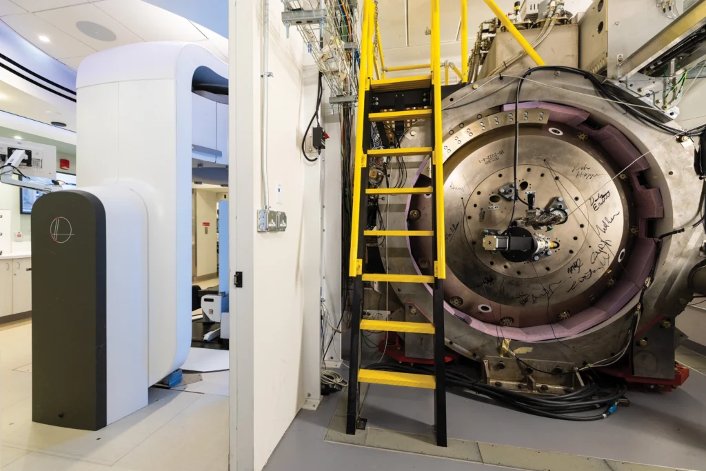
Protons are the positively charged particles in the center of an atom. Their charge and mass allow them to be maneuvered with precision. Especially for patients with tumors near critical structures — such as the brain, heart, spinal cord, or nerves involved in speaking and swallowing — protons offer advantages over traditional X-rays. Although they aren’t appropriate for all cancers, protons can be used for a wide variety of tumors, including those in the head and neck, spine, lungs, liver, and prostate.
“We can use magnets to send small bits of the beam of protons in various directions to shape the dose, even for tumors with irregular shapes,” said Yuan James Rao, MD, associate professor of radiation oncology and co-director of particle therapy. Protons can also be targeted to stop inside a tumor, with little to no exit dose. In contrast, X-rays travel through a tumor and keep going, exposing tissues behind it to radiation.
Smaller equipment expands proton therapy access
About four years ago, after failing to find a location in Palo Alto to build a traditional proton therapy facility, Loo and his team realized that they knew of two medical device companies taking different spins on smaller equipment.
Mevion Medical Systems offered the most compact cyclotron, which is the machine that generates the protons. These machines shrank proton therapy facilities to around half the size of a basketball court.
But the smaller cyclotron still required a three-story building. This was because, during treatment, patients would generally lie flat while the radiation source rotated on a gantry around them, moving one story above or below a treatment room.
Meanwhile, Leo Cancer Care took a different space-saving approach, developing a system that positioned patients upright for treatment in a specialized chair that swivels in the path of a stationary proton beam. “It’s much easier to rotate the patient, rather than having to rotate a big machine around them,” Loo said. His team wondered if the two innovations could be merged.
“With protons, we can deposit the dose of radiation in a more controlled way.”
Billy Loo, MD, PhD, professor of radiation oncology and co-director of particle therapy
“Once we brought the companies together, the light bulbs went on,” he said. Combining the world’s smallest cyclotron and the rotating chair would make a very small proton therapy setup, which could fit inside an existing 1,200-square-foot X-ray treatment vault in the Stanford Medicine Cancer Center. There would be no need to construct a new building. “They said, ‘OK, we will make this product,’ and Stanford Medicine agreed to be the first customer.”
The equipment makes proton therapy accessible to many Northern California patients who would have struggled to relocate hundreds of miles for the six to eight weeks often needed for treatment. Twelve other medical centers around the world are installing the new system.
Proton therapy research: upright treatment and FLASH
Loo and his team will conduct research on their new setup that includes testing the advantages of delivering radiation to patients sitting upright.
Evidence suggests that, for certain diseases such as lung cancer, sitting up may be safer — the lung is more stretched out and moves less when someone is upright than when they’re lying down, which should spare healthy tissue from radiation exposure.
Upright positioning also provides flexibility in how to deliver radiation. “It’s a new mindset of planning proton therapy using continuous arcs or even spirals of radiation, instead of beams from a few angles,” Loo said.
The Stanford Medicine team also plans to study an approach called FLASH treatment, giving the same dose of radiation in a much shorter time period than usual. Stanford Medicine experts began exploring this idea to improve the precision of dose delivery, because the body shifts even when someone is holding still.
While they were developing technology to deliver radiotherapy in a fraction of a second, researchers elsewhere demonstrated that very fast radiotherapy has the same impact on tumors but causes less damage to surrounding healthy tissues.
“It’s a really surprising scientific finding that we’re working hard to understand,” Loo said. “We plan to develop clinical trials of FLASH for our proton system.”
Pediatric radiotherapy that protects developing brains and bodies
The minimized side effects of proton therapy make an especially profound difference for children.
“Because kids are growing and developing, various parts of the body are more sensitive to even low doses of radiation,” said Susan Hiniker, MD, associate professor of radiation oncology and director of pediatric radiation oncology and associate director of particle therapy (pediatric) at Stanford Medicine Children’s Health.
Treatments for childhood cancers have vastly improved since Henry Kaplan’s day, giving survivors decades of life after cancer — and making it especially important to minimize their risk of long-term side effects.
In a serendipitous parallel to the experience of Kaplan’s team, Hiniker, Loo and their colleagues realized this spring that the best candidate for their first proton therapy patient was a young boy who could benefit from a novel, minimally invasive treatment.
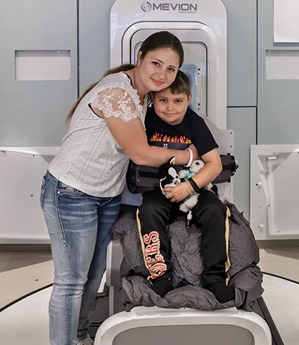
“He’s really excited about it. He’s been telling everybody, ‘I’m the first patient!’”
Tricia De La Torre; her son, Stephen, was Stanford Medicine’s first proton therapy patient
Seven-year-old Stephen De La Torre was diagnosed in early 2026 with an ultrarare brain tumor known as a papillary tumor of the pineal region.
The tumor, about the size of an AA battery, was blocking drainage of cerebrospinal fluid from his brain, causing swelling that led to headaches, vision problems, nausea and lethargy. On March 9, Stephen had surgery at Lucile Packard Children’s Hospital Stanford to excise the tumor.
“Our surgeons told us that his tumor was located really close to the brain stem, so they removed what they could safely,” said Stephen’s mom, Tricia De La Torre. “He needed radiation to eliminate the rest.”
When Stephen began radiotherapy treatment, the new proton therapy facility was in its final stages of calibration.
“Dr. Hiniker told us that he could receive protons for the last few radiation treatments, and that it would be good for the surrounding tissue not to get that extra radiation because the tumor is so close to his brain stem,” De La Torre said.
Stephen’s first proton treatment, which took about half an hour, was on June 4, three months after his surgery. He had six additional treatments in the following days, and in mid-June the team sent him home to Lakeport, California, where his family threw him a big party to celebrate his return.
Stephen was very brave about his entire medical journey, his mom said, adding that he kept reassuring his family he would be OK.
When it came to his role in Stanford Medicine’s tradition of innovation, “He’s really excited about it,” De La Torre said. “He’s been telling everybody, ‘I’m the first patient!’”
