
Computer memory
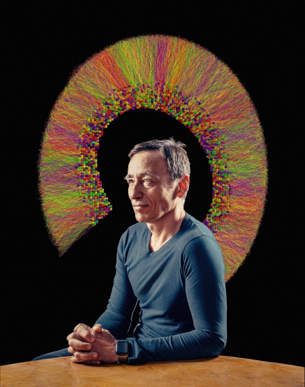
Capturing the brain’s learning and recall motor in silicon
In 1953, a 27-year-old Montreal man who’d had frequent seizures ever since getting a nasty smack in the head at age 7 underwent surgical removal of brain tissue containing the site where his seizures originated. The excised tissue included both of his hippocampi.
The hippocampus is a little horn-shaped structure found on each side of the brain’s midline just above the ears. If you spend enough time staring at a cross section of it on a slide, you may eventually come to see it as resembling a seahorse, which is what the Latin terms hippo and kampos roughly translate to.
In the 1950s, not much was known about the role of the hippocampus — or any other brain structure. The plight of the patient, Henry Molaison — referred to as H.M. — is widely known among neuroscientists as both a cautionary tale and a wake-up call.
Sure enough, his seizures did subside. But for the rest of his long life (he died in 2008 at 82), he could not remember anything new — not a single thing — for more than 30 seconds. His pre-existing biographical memories were unaffected.
“You could have a perfectly lucid conversation with him, walk out of the room to get coffee, come back in and have the same exact conversation all over again,” said Ivan Soltesz, PhD, professor and vice chair of neurosurgery at the Stanford School of Medicine. “For H.M., it was always as if for the first time.”
H.M. could learn new motor skills just fine but couldn’t remember learning them. Plus, his spatial memory was shot. He couldn’t get around on his own.
From his experience and many thousands of unrelated experiments, brain scientists and brain surgeons have learned that the hippocampus is both indispensable for learning and memory and, often, the seat where epileptic seizures are initiated.
These features, along with some anatomical and physiological ones that make it easy and exciting to study, have propelled the once-mysterious hippocampus to the fore as arguably the most thoroughly researched part of the brain. Much of this learning has come about by carefully taking the hippocampus apart and analyzing the activities of its components and connections.
But Soltesz, who has devoted more than 30 years to understanding how brain circuits work (or don’t), has gone a step further, taking to heart a thought attributed to the late, famed Caltech physicist and Nobel laureate Richard Feynman: If you can build it, you can understand it.
“If we’re telling the computer that ‘the firing frequency, strength and duration of this neuron-to-neuron connection should be this much,’ it’s because that’s what we’ve observed in biological systems. We don’t make stuff up.”
Soltesz, Stanford Medicine James R. Doty Professor of Neurosurgery and Neuroscience, and his teammates are building a full-scale virtual model of the hippocampus. But unlike a wooden airplane you might see hanging from a 12-year-old’s bedroom ceiling, this model lives in a computer, in the form of mathematical depictions of neuronal types and their electrochemical components and connections that drive the reception, propagation and handoff of nerve impulses.
The resulting mathematical constructs mimic the component processes that go into a neuron firing off an impulse or its failure to fire one. As a result, the properties of any given individual virtual neuron, or its connections with other virtual neurons, are very similar to what you’d find in its biological counterpart.
“Anything we feed into the model is based on hard experimental evidence,” Soltesz said. “If we’re telling the computer that ‘the firing frequency, strength and duration of this neuron-to-neuron connection should be this much,’ it’s because that’s what we’ve observed in biological systems. We don’t make stuff up.”
They’ve completed two of its most important sections — about half of the entire structure — and used them to illustrate what goes on inside their flesh-and-blood counterparts. The project, funded by the National Institutes of Health and the National Science Foundation, is steaming along, with plans to complete the model within two or three years.
That, in turn, will enable neuroscientists to get a better handle on how the hippocampus does the immensely important things it does: namely, ruling over two crucial cognitive processes. The first is episodic memory (what I had for breakfast), and the second is spatial memory (where I parked the car).
The ability to run virtual experiments may also speed the much-sought understanding of why the hippocampus is so vulnerable to normal aging — leading us to forget what we ate for breakfast and where we parked the car — and is so particularly prone to the biological deterioration set in motion by Alzheimer’s disease.
It may even lead to better understandings and treatments of a wide range of neurological conditions. And, of course, it’s the next step to the logical final outcome: a virtual brain.
Replicating anatomy
Modeling the hippocampus at such a level of biological realism is a tall order. Like all brain regions, it’s mostly a complicated tangle of individual neurons. The average neuron in the hippocampus is in communication with something like 1,000 other neurons, so the circuit diagram quickly gets crazy complicated. Making matters more daunting, not all neurons are alike.
About 80 percent of hippocampal neurons are excitatory: Their output signals have a stimulating effect on neurons they contact. Neuroscientists have divided the hippocampus into four or five serial compartments whose boundaries are roughly defined by where each compartment’s excitatory neurons pass along information to their downstream partners. The first of these compartments is called the dentate gyrus, which plays a crucial role in the transfer of information. That’s followed by compartments known as CA3, CA2 and CA1. (CA2 is very small and somewhat underexplored.)
Most of the remaining neurons in the hippocampus are inhibitory, with their outputs exerting an impulse-stifling effect on downstream neurons. Unlike excitatory neurons, their reach is almost entirely restricted to other neurons within their own compartments. So, the hippocampal inhibitory neurons are also known as interneurons. An interneuron can impinge on both excitatory neurons and other inhibitory interneurons, forcing anyone trying to parse the logic of compartmental circuitry to cope with cascades of double and triple negatives.
“It would be valuable, from both medical and strict scientific standpoints, to know what will happen if — say, as a result of illness or trauma — the hippocampus loses 20 percent of one or another particular population of neurons,” said Soltesz. But in the majority of cases, there’s no known way to experimentally manipulate the activity of a specific neuronal subtype.
“That’s where we come in,” said Soltesz, referring to his virtual hippocampus.
Exploiting the model
In 2017, the Soltesz lab published a report in eLife on its virtual model of a rat’s hippocampal compartment CA1 replete with 338,740 neurons, most of them excitatory. The minority of inhibitory interneurons are crucial, though. Of the many actual interneuronal subtypes known to inhabit CA1 (they differ in how hard and how quickly they inhibit and which part of their target neurons they contact), the model captures the majority of those whose characteristics and connections are well-known. The model features an astounding 5.7 billion neuron-to-neuron connections.
The numbers involved are so gigantic that a four-second simulation of CA1 activity takes four hours to run on Blue Waters, a powerful supercomputer hosted by the University of Illinois at Urbana-Champaign. Blue Waters’ operating speed is measured in the number of mathematical calculations it can perform in one second: 10 to the 15th power, the equivalent of stringing a few hundred thousand high-performance laptops together to work in tandem.
Modeling the functions of a human hippocampus — 100 times bigger, with an accordingly astronomical number of neurons and connections among them — would crash any existing computer, however super. Besides, there’s much more experimental data available about rodents because you can do experiments with them that can’t be done on humans.
Soltesz’s CA1 model exists in isolation. But he and his colleagues have approximated the placement, strength and timing of the inputs that this compartment’s real-life biological counterpart would have received from an estimated 454,700 neurons coming into it from elsewhere.
Intriguingly, the researchers have found that this model spontaneously reproduces some important rhythmic firing patterns seen in real neurons in CA1. Soltesz thinks different phases of these rhythms serve a multiplexing function, like different channels on a TV, allowing separate streams of information to be processed in parallel and then routed to the right destination. When the timing is off, information is misrouted or loses its meaning. Researchers can maximize these rhythms by keeping the total level of input stimulation within a certain range.
Among the most prominent rhythms in the brain are so-called theta waves, which arise in the hippocampus. These occur when a mammal (including the human kind) is learning, moving around or dreaming. It turns out that the peak incoming-stimulation level for optimizing theta rhythms in Soltesz’s CA1 model duplicates what’s been determined using an isolated rat CA1 in a dish.
“The result we got almost numerically mimicked what happens in the biological brain,” Soltesz said. “It’s mind-blowing.”
Soltesz’s team went beyond merely recapitulating experimental findings and performed some simulations whose outcomes couldn’t be predicted based on previous experimental experience. In their model, for instance, several different types of inhibitory cells needed to be transmitting impulses to recipient excitatory neurons at just the right time, frequency, speed and power or else the theta rhythm would collapse.
This finding doesn’t prove that the same thing would happen in a living animal’s hippocampus, but it generates a hypothesis that can in some cases be tested.
“I don’t care if the model’s predictions are proved true or false,” Soltesz said. “The point is to make experimentally testable predictions. The model is a tool, not a be-all and end-all.”
Playing around with an early, much simpler 10,000-cell model of another hippocampal compartment, the dentate gyrus, generated an intriguing hypothesis that not only was borne out but also could have huge implications for people with epileptic seizures.
“The result we got almost numerically mimicked what happens in the biological brain. It’s mind-blowing.”
“We became intrigued by a small population of cells in the dentate gyrus, called mossy cells,” said Soltesz. If neurons were airports, mossy cells would be JFK or LAX, he said. “These mossy neurons were especially hublike — each connects with many thousands of other neurons in the dentate gyrus.” His team wondered if the loss of some significant fraction of these sparse but densely connected neurons might increase the likelihood of symptomatic epileptic seizures.
Because mossy cells, which are inhibitory, hook up with both excitatory and other inhibitory neurons, trying to predict what will happen when you silence some of them is a toss-up. In a study published in Science in early 2018, Soltesz’s group succeeded in getting a clear answer. Their experiments in live mice showed that a die-off of mossy cells did trigger a rise in the spread of seizures originating in the dentate gyrus throughout the mice’s brains. It also reduced the mice’s ability to recall spatial information.
Mossy cells are especially delicate and vulnerable to pressure as well as to decreased oxygen supply, which occur in brain injury or stroke. Concussions and stroke are known risk factors for increased seizure susceptibility. A drug aimed at protecting mossy cells could have major clinical possibilities.
Soltesz’s models, which he makes freely available, have been used by labs all over the world for research on memory storage and retrieval, antiepileptic drug effects, genetic models of disease and more. He makes available all the experimental data his team has fed into the model, so the data can be funneled into any new, improved model that comes along. He’s even giving away software to investigators inclined to tinker with the model.
“What’s most impressive about Ivan’s CA1 model,” said Angus Silver, PhD, a professor of neuroscience at University College London and a Fellow of the Royal Society, “is that it’s full-scale. He and his team have combed the literature for all the information available about this part of the hippocampus, they’ve put it all together and, amazingly, it exhibits several key properties of the real CA1, without your having to tweak it. It certainly gives you hope that this approach is useful.”
Meanwhile, Soltesz and his team are forging ahead with a full-scale model of the rodent dentate gyrus (1 million cells in all, three times as many as in CA1), which Soltesz expects to complete in less than a year. Next on the to-do list are CA3, then models of nearby structures that send inputs to the hippocampus. In the longer term, he wants to model the entire brain region that includes the hippocampus — the temporal lobe.
But why stop there? Other well-funded groups, such as the Allen Institute for Brain Science in Seattle and the Human Brain Project in Switzerland, are also building representations of various parts of the brain.
“We’re not working in a vacuum,” said Soltesz. “I can imagine that in 20 years or so we’ll have full-scale models of mouse and human brains at single-cell resolution.” It may someday be possible, Soltesz speculated, to build customized, patient-specific models of the entire brain based on advanced noninvasive imaging and recording techniques, so the effects on any patient’s brain of a given drug or other intervention can be tested.
When a model has as much resolution as the thing it’s modeling, is it still a model? Or is it a working copy?
“That’s an excellent question,” Soltesz said. “Luckily, we’re nowhere near being able to answer it. It’s pure fantasy right now. But we humans are busily working our way toward that quandary. We’ll deal with it when we get there.”
