
Opening stroke’s window
Treating stroke more than a few hours after the crisis is no longer considered absurd
Around 10:30 p.m. on April 23, 2017, Cindi Dodd ended a phone call with her best friend, turned off her bedside light, and settled in for a night’s sleep in her Salinas, California, home ahead of a surgery the next day.
The scheduled operation was a breast reconstruction following a bout with cancer, but when Dodd’s husband, Rick, woke her at 5 a.m., something else was terribly wrong. She seemed to be having a seizure. She couldn’t move her left arm or leg, and when she spoke, only gibberish came out.
Rick Dodd called 911 and yelled to their son to turn on the porch light for an ambulance. At the hospital, the family found out that a blood clot had lodged in Cindi Dodd’s middle cerebral artery, preventing blood and nutrients from flowing to her brain. While she was asleep, the 46-year-old graphic designer had experienced an acute ischemic stroke. Cells in her brain had begun to die.
Doctors told Dodd’s family they could do nothing to remove the blockage: Too much time had lapsed since she was last known to be well. According to standard guidelines, after six hours from the onset of a stroke, the risk that treatment would cause dangerous bleeding in the brain and elsewhere in the body outweighed the benefits of restoring blood flow to damaged tissue.
Her best option, an emergency physician told her husband, was to get to Stanford Health Care. There, doctors were leading a large clinical trial to determine if the narrow window of time for stroke treatment could be widened for patients under some circumstances.
Rick Dodd didn’t hesitate. Within an hour, a helicopter arrived, and Cindi Dodd was on her way to Stanford Hospital.
“That’s my baby. Take care of her,” Dodd’s mother told the flight nurse through tears.
Extending treatment timeline
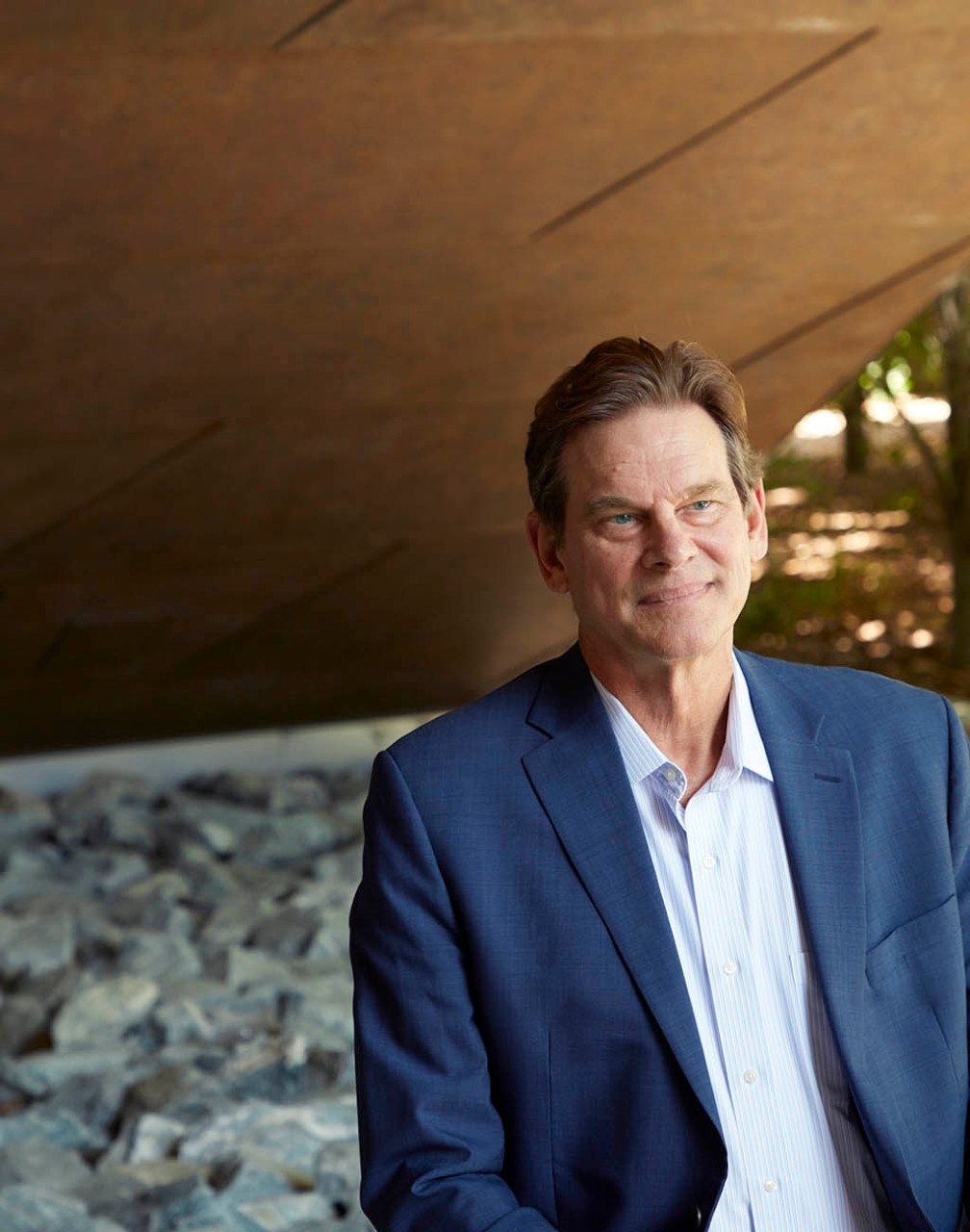
On the other side of that flight, at Stanford Hospital, Gregory Albers, MD, was leading the most critical clinical trial of his 30-year career.
He became fascinated with the human brain when he was a teenager, in California’s San Fernando Valley. Since completing a stroke research fellowship at the Stanford University School of Medicine in 1988, he had dedicated his work to improving the lives of stroke patients.
Whenever stroke patients arrived at the hospital too late for a dismal outcome to be prevented, Albers always felt dismayed. He wondered if some patients who arrived many hours after their stroke began might still benefit if blood flow was restored to their brain tissue. And if so, was there a way to identify which patients could be helped during this longer period?
These questions would frame his professional efforts for nearly three decades, beginning in the 1990s.
During that time, through a methodical progression of research, Albers and his colleagues chipped away at the status quo of stroke treatment. They tested — then refined — a new imaging technique that could quickly reveal the damage wrought by a stroke, as well as the at-risk tissue that could still be saved.
Gradually winning over skeptics, they crafted a protocol that added precious hours to the timeline for stroke care and carried immense potential to help countless future patients avoid severe disability or worse.
“It was a huge challenge,” Albers said of his quest to improve outcomes for stroke patients. “We were dealing with the No. 1 cause of disability worldwide, and there was no treatment.”
Advancing brain imaging
The Stanford Stroke Center opened in 1992, led by Albers, neuroradiologist Michael Marks, MD, and neurosurgeon/neuroscientist Gary Steinberg, MD, PhD. The center’s aim was to develop new therapies and bring them to patients.
“We were not satisfied with this nihilistic approach, that stroke is something you can never treat,” said Steinberg, who is the Bernard and Ronni Lacroute-William Randolph Hearst Professor in Neurosurgery and Neurosciences.
Within a year of establishing the center, its leaders were approached by Michael Moseley, PhD, a physicist who had recently joined Stanford Medicine’s radiology department. Moseley was studying ways to capture images of brain damage from a stroke, and he’d made an intriguing discovery.
At the time, because of limitations of conventional magnetic resonance imaging and computed tomography, evidence of injury from a stroke didn’t appear on a scan until four or more hours after a patient’s symptoms began.
“It was a huge challenge. We were dealing with the No. 1 cause of disability worldwide, and there was no treatment.”
Gregory Albers, MD, professor of neurosurgery
When brain cells begin to die during a stroke, they lose the ability to maintain a balance of water inside and outside the cell. Conventional techniques could capture images of dead cells swamped with water hours later, but Moseley wanted an earlier indication of what was happening.
In a late-night laboratory insight, he discovered that he could track a stroke’s damage to brain tissue as it happened through an MRI technique that was more sensitive to the movement of water in cells. Scans created through diffusion-weighted imaging showed a clear difference between cells that had no active transport of water and those with normal water-shuttling activities.
In short, Moseley had found a way to watch brain cells die in real time.
To Albers, “It seemed like the holy grail.”
With Moseley, Marks and other Stanford colleagues, Albers embarked upon a series of studies that used diffusion-weighted imaging to track stroke patients for weeks. The resulting images — with bright white smudges showing dead tissue, in contrast to healthy gray tissue — depicted a process that happened at different rates for different people, but often took more than a day to reach maximum size.
This discovery ran counter to the prevailing belief that a stroke completed its path of destruction within one to two hours.
Watching the progression of images brought the researchers to the next logical step: “I could see dead brain, but I couldn’t predict what was destined to die,” Moseley, a professor of radiology, said.
They decided to follow the blood. Using a technique called perfusion-weighted imaging, they injected a contrast agent into a patient’s bloodstream and monitored how long it took the contrast to reach distinct areas of the brain. If a major blood vessel was blocked, the contrast agent’s journey to that side of the brain was delayed.
It was hard to tell from a brightly colored perfusion-weighted image whether tissue was dying or already dead.
However, when the researchers compared it with a diffusion-weighted image, which clearly depicted dead tissue, they could determine whether there was a mismatch area — brain tissue that had not died but was marked for demise if the artery’s blockage wasn’t cleared.
They realized they could determine whether a person’s stroke was finished. If it was ongoing, they could predict how it would play out if physicians couldn’t open the blood vessel.
Their rough data suggested that about three-quarters of stroke patients could still benefit from treatment after six hours; after 12 hours, treatment could still help about half of them.
This was radically different from what animal models had shown: In rats, a stroke would typically complete within 90 minutes. In monkeys, three hours was the upper limit for avoiding brain damage, according to a pivotal study.
In their 1999 paper, Albers and his colleagues predicted that late-window therapy could improve stroke outcomes, but their study was met with ambivalence from other neurologists. Some were intrigued, while many others thought this approach had no chance of success.
“Neurologists in general tend to be skeptical people,” said Albers, the Coyote Foundation Professor, “and there had been a number of stroke trials in the past — many, many stroke trials that had been done in the ’80s and ’90s — that neurologists were initially excited about. They all failed.”
A promising new drug
Before the 1990s, physicians routinely let a patient’s stroke run its course, convinced there was nothing they could do. This changed as scientists gained a better understanding of the rate at which human brain cells disintegrate and as doctors tested tools for restoring blood flow to oxygen-starved tissue.
A clot-dissolving drug called tissue plasminogen activator — known as tPA — showed promise. It works by activating plasmin, an enzyme responsible for breaking down blood clots. However, because tPA can increase the risk of bleeding everywhere in the body, it can cause serious bleeding complications.
In 1996, the U.S. Food and Drug Administration approved tPA for treating ischemic strokes — those caused by a blocked artery — but only within three hours of the first symptoms. After three hours, the thinking went, it was too late to save the dying brain tissue, and the risk of the drug causing further harm outweighed any potential benefit.
Because many patients are unable to seek medical care immediately, the time constraint strictly limited the number of patients who could be treated with tPA.
Albers believed more people could be helped.
In 2001, after he, Moseley and their team published several preliminary studies about the new imaging technique, they were funded by the National Institutes of Health to perform a study of stroke patients who had not arrived in time to receive tPA. The patients would undergo the new imaging techniques and be given tPA between three and six hours after their first stroke symptoms.
“Neurologists in general tend to be skeptical people and there had been a number of stroke trials in the past … that neurologists were initially excited about. They all failed.”
Gregory Albers
Because tPA works for only about half of the patients who receive it, that meant the researchers could test their imaging technique in two ways. For patients with a mismatch and whose blockage persisted, they could see whether the stroke proceeded as predicted. For patients with restored blood flow, they could see if the intervention prevented damage in the brain areas predicted to be salvageable, and if those patients had better outcomes.
The results, published in 2006 as the Diffusion and Perfusion Imaging Evaluation for Understanding Stroke Evolution (DEFUSE) study, turned out exactly as Albers and the Stanford team had hoped and predicted.
Patients with a mismatch whose arterial blockage was dissolved showed improvement in functioning, such as regaining control of their limbs, as measured on the NIH Stroke Scale. This suggested that the imaging had successfully identified salvageable tissue in individual patients and that restoring blood flow by administering tPA had saved the tissue.
What’s more, the study indicated that more than half of the patients still had brain cells that could be saved five to six hours after a stroke.
“Clearly,” Albers thought, “we’re on to something here.”
Expanding the limits
His team’s next study pushed the envelope further.
Though the design of the second DEFUSE trial was similar to the first, there were two crucial differences. First, the time window was doubled to 12 hours. Second, patients would be treated with a new technique called thrombectomy, in which a physician threads a catheter through the artery to the clot, then deploys a mechanical device to extract it.
Additionally, the team had developed an imaging software called RAPID that automatically analyzed the diffusion and perfusion images and calculated the volume of salvageable tissue; the nine medical centers participating in the study all used the new software in the trial.
Some of the researchers’ peers expressed doubts about the study’s premise — 12 hours was so much longer than the accepted window for stroke treatment. But when the second DEFUSE study was published in 2012, six years after the first, the findings were undeniable.
“It worked beautifully,” Albers said.
Their results suggested that the window for successful treatment could be extended to 12 hours for some stroke patients. But they had not yet conclusively shown that patients selected with their imaging technique benefited from late treatment. Doing so required a randomized trial, which is what Albers wanted to do next.

‘Brain is of the essence’
In theory, thrombectomy devices, with their capability to clear blockages, were the perfect instrument to help physicians save brain tissue in stroke patients. However, studies of early versions showed disappointing results. Although the devices could reliably restore blood flow to oxygen-deprived brain tissue, stroke patients’ ability to function often didn’t improve much.
In the years after the DEFUSE 2 study was published in 2012, manufacturers improved the devices. Meanwhile, Albers wondered if many of the patients being treated already had irreversible injury. He wanted to find out if patients who underwent a thrombectomy fared better when the Stanford team’s imaging had shown salvageable tissue prior to the procedure.
Before the team could secure funding for a third DEFUSE trial, however, several studies were published that seemed to confirm Albers’ theory, widening the stroke treatment window to six hours in certain cases. Two had used the RAPID software, which was now owned by a company Albers had co-founded, and these studies produced the greatest treatment benefits.
Among patients with scans that showed salvageable tissue, those who received a thrombectomy within six hours of a stroke showed better outcomes than those who had not undergone the procedure. In 2015, the American Heart Association/American Stroke Association changed treatment guidelines to reflect what the studies had found.
But the Stanford researchers did not believe the issue was settled, as six hours was not long enough to get to the majority of stroke patients; many live far from a stroke center or have the stroke while they’re sleeping.
“They could have said, ‘You don’t need any more patients. You’ve proven what you need to prove.’ But they also could have said, ‘Oh, no, you’re not there yet, and you’re not going to get there.’”
Stephanie Kemp, the Stanford Stroke Center’s program manager
The Stanford studies suggested that for patients flagged through imaging, treatment could succeed up to 24 hours after symptoms began. The team was finding that “time is not of the essence. Brain is of the essence,” said Maarten Lansberg, MD, PhD, a Stanford professor of neurology who joined the effort in 1997.
The third DEFUSE trial, also funded by the NIH, began in 2016 with more than three dozen medical centers participating. The randomized study focused on patients whose stroke was caused by a blood clot obstructing one of two large arteries in the brain — the middle cerebral artery or the internal carotid artery. This happens in about 1 in 4 ischemic strokes and accounts for the most disabling strokes.
If the RAPID software showed potentially salvageable brain tissue six to 16 hours after a stroke’s onset, a patient was randomly assigned to have either standard medical treatment or an alternative that included both the standard treatment and a thrombectomy. Participants were tracked for three months, the period when stroke patients typically experience most of their recovery.
Meanwhile, another study using the Stanford-designed software had been launched in 2014 by Stryker Corp., which manufactures devices for stroke care. The design of Stryker’s trial, called the DAWN study, was almost identical to the DEFUSE 3 study with one significant difference: It pushed the treatment window to 24 hours after a stroke’s onset, compared with 16 hours in DEFUSE 3.
After an analysis of interim data, the data safety and monitoring committee stopped enrollment in the DAWN trial early, in February 2017. Among 206 stroke patients whose scans showed salvageable brain, those who underwent a thrombectomy experienced less disability than the control group. Because the DEFUSE 3 study was so similar, the NIH placed it on hold soon afterward, in June 2017, to evaluate preliminary findings.
Those weeks were nerve-wracking for the Stanford team, who waited to see what would come next for the study they’d worked so long to begin.
“We were nervous,” said Stephanie Kemp, the Stanford Stroke Center’s program manager. “They could have said, ‘You don’t need any more patients. You’ve proven what you need to prove.’ But they also could have said, ‘Oh, no, you’re not there yet, and you’re not going to get there.’”
Changes in treatment guidelines
In fall 2017, when Albers finally saw the data from 182 participants in the Stanford-led trial, he couldn’t sleep.
There was no need to restart enrollment. The data showed that the imaging technique Albers and his colleagues had refined over so many years could help physicians determine when they could do more for a stroke patient and substantially improve the patient’s recovery.
Three months after a stroke, 45% of the patients who received a thrombectomy six to 16 hours after their first symptom were functionally independent, compared with 17% who received standard care. Among patients receiving the thrombectomy, 14% died within three months of having a stroke, compared with 26% in the control group. The team’s findings underscored the results of the DAWN trial.
Albers thought back across the decades — his entire career. He thought about the times he’d had to tell patients, “I’m sorry. You came in too late. We can’t treat you.” About how, for some future patients, the likelihood of death and disability was now cut in half.
“This is going to change the world,” he thought.
The publication of the DEFUSE 3 study in the New England Journal of Medicine was timed to coincide with the American Heart Association’s International Stroke Conference in January 2018. On the day Albers presented the results of DEFUSE 3, the association announced changes to treatment guidelines for acute ischemic stroke, recommending a treatment window for mechanical clot removal up to 24 hours after onset in certain patients with clots in large vessels.
William J. Powers, MD, chair of the neurology department at the University of North Carolina at Chapel Hill at the time, headed the committee in charge of recommending guidelines. He knew that many researchers had conducted similar investigations, but the Stanford team’s contribution stood out.
“There’s plenty of papers out there that say, ‘Oh, we think we can, with 60% or 80% accuracy, identify this tissue that will live or die,’” Powers said. “But what distinguishes them — and what they really should get credit for — is being the ones who came up with a practical way to do this. They took it on, did the clinical trial, and proved that it worked.”
Powers said he has integrated the approach into his own practice, as have untold numbers of health care providers. As of July 2021, the software originally developed at Stanford was being used in more than 1,800 medical centers around the world, and more than 2 million scans had been performed. At Stanford Health Care, about five stroke patients undergo a mechanical thrombectomy after perfusion imaging every week.
‘A fighting chance’
After Cindi Dodd arrived at Stanford Hospital on that April day in 2017, a CT scan showed salvageable tissue in her brain. As part of the DEFUSE 3 clinical trial, Dodd underwent a thrombectomy to remove the clot blocking her artery.
When she woke up in the intensive care unit, her family told her what had happened. Her first thought was that she was too young for a life-threatening health scare. Then she realized the significance of the clinical trial: “It gave me the opportunity to fight for my life.”
Dodd’s rehabilitation was gradual, but after a year, she was back to walking, talking, driving and working.
“I thank you for your study that gave me a fighting chance at living as a functional human being, a contributing member of society! I will forever be thankful for you.”
Stroke patient Cindi Dodd
She never met Albers in person, but on Thanksgiving Day 2018, Dodd looked up his email address and started to type. She told him about herself, that she was a wife with dreams of traveling and a mother with a fierce desire to be there when her two children graduated from college, married and had children of their own.
“I thank you for your study that gave me a fighting chance at living as a functional human being, a contributing member of society!” she wrote. “I will forever be thankful for you.”
The next day, Albers replied.
“It has been such an amazing year for our group to see the dream that we have had for two decades finally come true,” he wrote. “We are so grateful to patients like you, who were willing to take a chance on a new approach to treating stroke.”
— Contact Amy Jeter Hansen at ajeterhansen@stanford.edu
