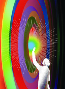
Dangerous infiltrators
Cancer hijacks nervous-system signals — and scientists want to stop it
About 15 years ago, Stanford Medicine neuro-oncologist Michelle Monje, MD, PhD, began to suspect that the brain tumors she studied were doing something strange. Cancer cells sometimes copycat their healthy counterparts, so Monje and her team weren’t surprised to uncover simple parallels between healthy and malignant brain cells. The cancer’s biological “borrowing” was similar to a symphony-goer who whistles the theme from a concerto on the bus ride home.
But the team’s data hinted that these brain tumors were orchestrating something much more complex. Instead of just humming the themes of healthy brain biology, the research suggested the tumors could round up many important cell-signaling instruments — the microscopic equivalents of, say, violins, cellos, flutes and trombones — and use them to play a score of its own.
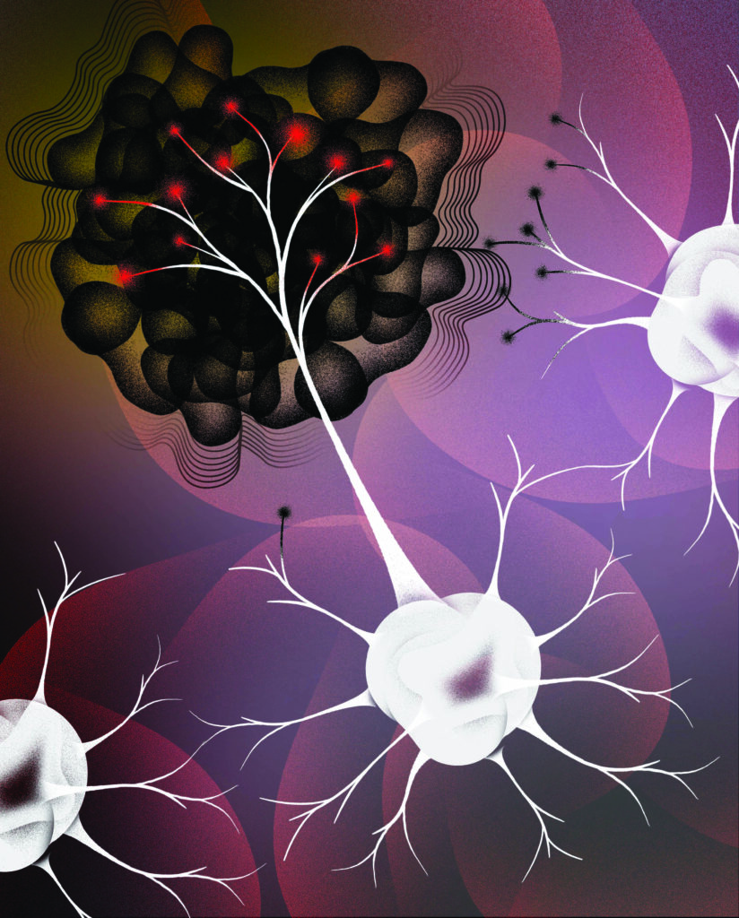
In physiologic terms, Monje’s team gradually demonstrated, certain cancer cells form working electrical connections with nearby nerves. The tumors wire themselves neatly into the brain’s electrical apparatus, then use healthy nerves’ signals for their own purposes — to drive malignant growth. These cancers also hijack the machinery of learning to strengthen connections with the healthy brain and further enhance their ability to multiply.
Her team’s findings form the foundations of the medical field called cancer neuroscience. It offers opportunities to target some of the worst forms of cancer, including brain tumors that are almost always lethal. Scientists are especially intrigued by the cancer treatment potential of drugs approved by the Food and Drug Administration for other neurologic disorders, such as epilepsy. It turns out that several such medications interrupt neural signals now understood to drive certain cancers.
Clues about a terrible tumor
Many of the Monje lab’s early insights came from a tumor called diffuse intrinsic pontine glioma, a cancer that causes its young patients to experience profound debility — unable to walk, eat or hear, for example — on the way to dying from their disease, often within a year of diagnosis.
DIPG cells originate in the brain stem, a region that controls essential body functions such as breathing and heartbeat. They entwine with healthy cells, which makes surgical removal impossible. The five-year survival rate is less than 1%.
When she saw DIPG co-opting large portions of normal neurobiology, Monje’s thoughts went first to the kids and teens she has treated.
“It was so striking: As a clinician, I felt that suddenly a lot of things make sense,” she said. For one thing, the discoveries explained why the tumors spread like wildfire through the brain and spinal cord but didn’t become established elsewhere in the body.
Even better, the knowledge offered hope for new treatments.
“Seeing this cancer structurally integrating into neural circuits, I thought, ‘Of course it has been intractable. Now we have to approach this disease in a very different way; it’s really a disease of neuroscience.”
Michelle Monje, MD, PhD
“Seeing this cancer structurally integrating into neural circuits, I thought, ‘Of course it has been intractable,’” said Monje, the Milan Gambhir Professor in Pediatric Neuro-Oncology. “Now we have to approach this disease in a very different way; it’s really a disease of neuroscience.”
Monje’s work is breaking a logjam in the understanding of DIPG and other brain and spinal cord tumors in the same family, known as high-grade gliomas for their rate of growth and the cells from which they originate.
For decades, no one studied the biology of DIPG. Surgeons rarely took biopsies of the tumors because of their sensitive locations. This meant the cancer cells weren’t available for scientists to study. In 2009, Monje began asking families whose children were dying of DIPG if they would be willing to donate postmortem tissue samples for research. At first, she was nervous about asking, but she found that many families welcomed an opportunity to help scientists understand the disease that was taking their children from them.
Thanks to the tumor tissue samples — more than 110 donations to date — the research team has been able to grow the tumor cells in the lab, implant them in mice, share these models with other teams around the world and make a number of important advances.
“The cancer is diffusely and widely invading the nervous system because that’s advantageous for it,” Monje said.
Hardwired into the brain
Once Monje began to suspect that DIPG forms electrical connections with nearby brain cells, she asked Robert Malenka, MD, PhD, to help her team test it. He has four decades of experience studying neurons’ electrical connections, known as synapses.
“We thought this work was going to take two months, and it ended up being a several-year, very difficult project,” said Malenka, the Nancy Friend Pritzker Professor in Psychiatry and the Behavioral Sciences.
For one experiment, a scientist from Malenka’s lab, Wade Morishita, PhD, set up a way to listen for electrical activity in one tumor cell at a time. It was a key step in proving that the tumors form real synapses that receive signals from their healthy neighbors. The project was part of a groundbreaking study led by Humsa Venkatesh, PhD, then a graduate student mentored by Monje, which was published in 2019 in Nature.
Discovering that the tumors wire themselves into the brain was “unsettling,” Monje said at the time, adding, “This is such an insidious group of tumors. They’re actually integrating into the brain.”
“We thought this work was going to take two months, and it ended up being a several-year, very difficult project.”
Robert Malenka, MD, PhD
Via synapses and additional electrical connections between adjacent cells called gap junctions, about half of all glioma cells in a given tumor have some type of electrical response to signals from healthy neurons, the research showed.
The discovery that cancer cells form working synapses shocked experts, Malenka said. “It looks like normal synaptic communication, and that was very surprising,” he said. “To be honest, I wouldn’t have predicted it.”
Malenka was surprised because the brain is extremely picky about where it makes new connections. The brain’s ability to selectively build and get rid of synapses is a major aspect of learning.
“It’s not as if each nerve cell automatically makes synapses with every other nerve cell in its vicinity,” Malenka said.
“It’s highly orchestrated. Now you’re throwing in this bizarre group of cancer cells? You wouldn’t want them to incorporate.”
In addition to communicating via synaptic connections, healthy brain cells also signal to each other with protein messengers that move between cells to trigger complex intracellular responses. Such responses include molecular signals that underlie the neural plasticity needed for learning and memory. (The brain physically changes as we learn; these signals are part of that change.)
Investigations over the last several years by Monje’s team showed that tumors co-opt two of the messengers, proteins called neuroligin 3 and brain-derived neurotrophic factor, or BDNF, to strengthen their synaptic links to healthy cells.
“It’s the same electrical activity that helps us think, move, feel, touch and see. Cancer is plugging into that and using that to grow, invade and even occur in the first place.”
Kathryn Taylor, PhD
During the studies of BDNF, which were led by Kathryn Taylor, PhD, then a postdoctoral scholar in the Monje lab, and published in Nature in 2023, one key experiment showed that when the cell machinery triggered by BDNF was activated more, the tumor cells responded with stronger electrical currents, which then fueled their growth. It was clear evidence that the cancer uses the brain’s learning machinery to grow.
“We looked at the electrophysiological recordings and seeing this increase was … I will never forget that. It was pretty incredible,” Taylor said. “What was so striking about that finding was that not only can the cells connect, they also dynamically respond to input from healthy brain cells. The tumor cell is not only plugging into the network, it’s increasing its connection to that plug.”
It’s disconcerting that tumors use brain activity to grow, Taylor admitted. “It’s the same electrical activity that helps us think, move, feel, touch and see,” she said. “Cancer is plugging into that and using that to grow, invade and even occur in the first place.”
Hope for treatment
But understanding these interactions between tumors and the healthy nervous system presents new options for cancer treatment. Through their research published in the 2023 Nature article, Taylor, Monje and their team showed that medications aimed at the BDNF receptor, developed for other forms of cancer, work surprisingly well at slowing the growth of DIPG and related neurologic tumors.
Other drugs, including certain painkillers, anti-seizure medications and blood pressure medications, also have potential as cancer fighters. A detailed understanding of how the tumors tap nerve signals to grow provides a huge leg up in cancer treatment research, as scientists can match what’s in the “medicine cabinet” of FDA-approved neuroactive drugs with their new knowledge of how cancers operate.
Stopping the worst gliomas, including DIPG, will require a mixture of tactics, from cancer neuroscience and from other oncology specialties, Monje said. Perhaps doctors can start treatment with neurological medications that slow the tumors’ growth, then give immunotherapies — such as specially engineered immune cells called CAR-T cells, which her team is also studying as a treatment for DIPG — as a second line of attack. Such a strategy might give immunotherapy treatments enough of a head start to enable them to outpace the rapidly growing tumors.
“The metaphor for DIPG that has been in my mind for several years now is that of a house fire,” Monje said. “The CAR-T cells are the firefighters, the mutation that caused the tumor was the spark that ignited it, and it’s like there’s a gas line open, pouring gas on the fire — those are the neuronal signals. The firefighters have a really tough job to do if the gas is still pouring in. We have to switch off the gas.”
Beyond the brain
Cancer neuroscience is also turning up clues for ways to tackle tumors that spend all or part of their time outside the brain. Nerves normally send signals to stem cells that help regulate healthy organ development and repair, and research is increasingly documenting that these signals can fuel cancer cells. “There are critically important roles for the nervous system in pancreas, prostate, breast, colon, gastric, skin, and head and neck cancers — a very long list,” Monje said.
Monje’s discoveries about brain tumors caught the attention of cancer biologist Julien Sage, PhD, professor of genetics and the Elaine and John Chambers Endowed Professor in Pediatric Cancer. Sage studies small cell lung cancer, an aggressive disease caused primarily by cigarette smoking.
Monje’s lab and offices are directly above Sage’s in the Lorry I. Lokey Stem Cell Research Building. Their conversations made him wonder if a neurobiological approach could help crack one of the hardest problems his team faces. Small cell lung cancer spreads readily to the brain, where its metastatic tendrils are often fatal.
“Treating brain metastases is a really, really challenging clinical problem,” Sage said. “Many drugs don’t go to the brain, and if you mess up with the brain, you mess up with the person.”
“Treating brain metastases is a really, really challenging clinical problem. Many drugs don’t go to the brain, and if you mess up with the brain, you mess up with the person.”
Julien Sage, PhD
Sage believes that small cell lung cancer cells invade the brain because they are equipped to take advantage of local conditions. “It would make sense that if you have a thousand cancer cells and they land in a thousand different places in the body — if they land in places where they have friends, they’re going to stay,” he said.
His team’s work, published in 2019 in eLife, supports the concept. The work was led by then-graduate student Dian Yang, PhD, now a faculty member at Columbia University, in a joint effort with the Stanford Medicine laboratory of Monte Winslow, PhD, associate professor of genetics and of pathology.
The researchers showed that the cancer cells become more nervelike in the brain. While growing in the lung, the cancer cells are small and round; in the brain, they extend protrusions that look like axons, Sage explained. (An axon is a neuron’s long, stringy arm through which an electrical signal can travel to reach the next cell that needs its message.) As they establish a metastasized tumor in the brain, small cell lung cancer cells engage in a strange new activity: “The cells are round, then they extend the axonlike protrusion and they migrate after their protrusion,” Sage said. “They use the protrusion as a way to grab onto something.”
It’s a behavior that neuroscientists recognize. “This is how the brain is formed,” Sage said. “These cancer cells look more like neurons during development.” The cancer cells also make neighboring brain cells look more like early-in-development versions of themselves. They “kind of reprogram the brain microenvironment,” Sage said.
The scientists hope that this reprogramming trick offers a toehold for treating small cell lung cancer in the brain. The disease occurs mostly in older adults, whose healthy brain cells are not developing. If researchers can identify drugs that target the development-like aspects of cancer, those drugs might kill the cancer without causing too much damage to the adult brain.
Sage’s team is extending these studies into a collaboration with the labs of Monje and Venkatesh, now an assistant professor at Harvard Medical School, to understand whether the lung cancer cells are able — as glioma cells are — to form synapses and receive electrical signals once the malignant cells take up residence in the brain.
Hope for the future
From its start in an odd bit of experimental data — those first hints that brain tumors were orchestrating their fate, not just humming along — the field of cancer neuroscience continues to grow. Its origins bring to mind a quote from biochemist and writer Isaac Asimov: “The most exciting phrase to hear in science, the one that heralds new discoveries, is not ‘Eureka!’ but ‘That’s funny …’”
For Monje, who was inspired to study severe childhood gliomas such as DIPG more than 20 years ago, at a time when the biology of the disease was completely unknown, the new options are heartening. The old way of trying to treat the deadly tumor — a sort of throw-the-spaghetti-at-the-wall approach, using drugs not fitted to how the tumor grows — is obsolete, she said.
“This is a connected tumor; it’s connecting to the entire nervous system. We have to disconnect it,” she said. “We understand enough about this disease now to have lots of really rational ways to try to fight it.”
Editor’s update: On March 5, 2025, the Lundbeck Foundation announced Monje would receive the Brain Prize 2025. The foundation awards the Brain Prize annually to honor groundbreaking advances in any area of brain science. The 2025 award recognizes Monje’s pioneering advances in cancer neuroscience. She shares the $1.4 million award with Frank Winkler, MD, PhD, of Heidelberg University, another cancer neuroscience expert.
