A delicate operation
Removing a tumor from deep in a 2-year-old’s brain
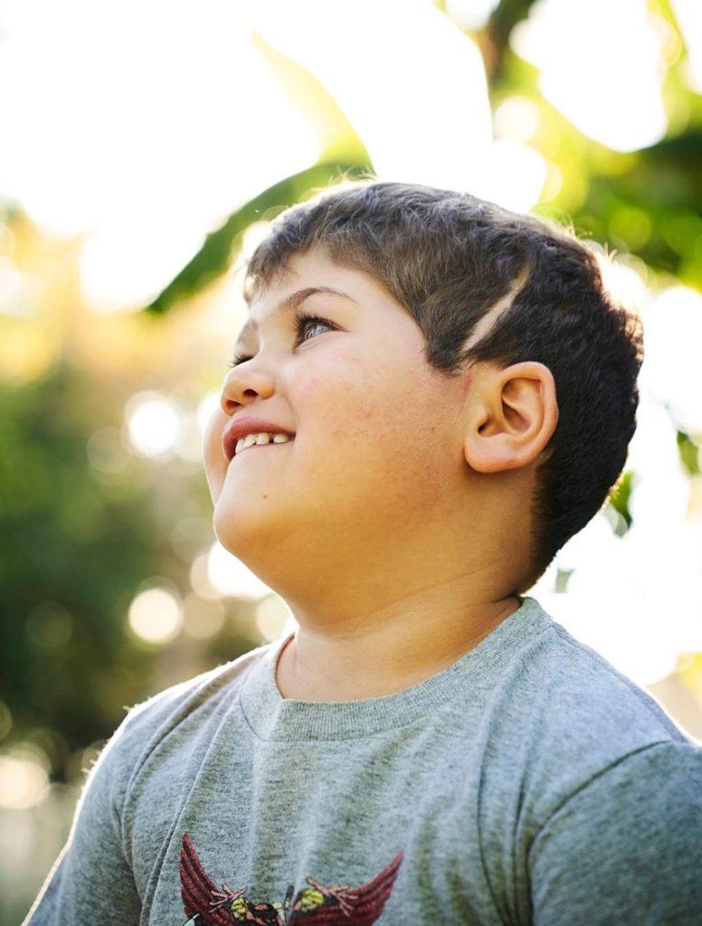
In August 2018, 2-year-old Ari Ellman’s parents took him to an emergency department near their home in San Francisco for the latest in a series of uncontrolled vomiting bouts. While awaiting an abdominal MRI, Ari had his first seizure, shifting doctors’ attention from his abdomen to his head.
A brain MRI revealed a golf-ball-sized growth in the difficult-to-reach central lower part of his brain, near the base of his skull. The rare, non-cancerous but fast-growing tumor, called a craniopharyngioma, was entangling the critical brain structures in the skull base. Unless the growth was removed, it would endanger all those structures and ultimately Ari’s life.
The Ellmans’ world turned on its head that day. “I barely had time to feel sorry for myself, though,” remembered Ari’s father, Jonathan. “A friend said, ‘There’s no time for self-pity, or anything else really … except focused action.’”
The Ellmans seized the reins of Ari’s care and didn’t let go. “The night after the diagnosis, my heart was all over the floor,” said Ari’s mother, Na’ama. “But Jonathan turned his computer toward me and said, ‘These are the top hospitals and craniopharyngioma surgeons we need to speak with. Tomorrow!’”
A daring approach
Millions of years of evolution buried such essential brain structures as the pituitary gland and hypothalamus in the bottom middle of the human head where they would be well-protected from a world full of sharp and heavy dangers.
So, it is no accident that the same important part of the brain, known as the skull base area, is notoriously difficult for surgeons to reach. For the first century of modern brain surgery, the only way to get there was by opening the top of the head, spreading the brain’s hemispheres apart, and tunneling down between them to the core.
Because the optic nerves, which connect the vision-processing part of the brain to the eyes, stand between the skull base area and that cranial opening, surgeons often had to work around those delicate and vulnerable structures, too.
Collateral damage to essential brain tissue on the way down could be devastating, and further damage could be imposed when surgeons were pulling a tumor up and out.
In the past decade, though, advances in imaging, surgical anatomy and surgical tools have enabled surgeons to use a less destructive approach. Instead of entering the skull from above, they enter through the nose and sinus area, just below the hardest-to-reach skull base structures.
This method, known as transnasal endoscopic skull base surgery, has become the preferred method for removing tumors in this part of the brain — but only in adults. Children have much smaller sinus cavities, and at the time Ari became ill, surgeons still approached pediatric skull base tumors the old-fashioned way — open surgery from above.
“A 2-year-old’s sinuses are only 20 millimeters wide or narrower. And you’re removing a tumor that may be wider than the nasal passage itself. It’s like getting a ship out of a bottle.”
Peter Hwang, MD, a professor of otolaryngology
However, as the Ellmans would soon discover, an extraordinary team of Stanford neurosurgeons and rhinologists believed a transnasal approach would be feasible even in small children. They just needed the right case to prove it.
If successful, they would not only have an opportunity to save the life of a dangerously ill patient but also to provide pediatric neurosurgeons with a technique for skull base surgeries in small children for generations to come.
Their method would give them direct access to essential parts of the young brain that have been excruciatingly hard to reach. A failure would make that path much more difficult for future surgeons to take — or even consider.
In the first week after learning Ari’s diagnosis, his parents sent his case to tumor boards — multidisciplinary groups of specialists — at 15 leading medical centers. Some suggested old-school open-brain surgery, which, in addition to the obstacles described above, often fails to remove the entire tumor, partly because the roots of craniopharyngiomas, at the bottom of the brain, may be inaccessible from above.
Other surgical groups suggested radiation, but that can cause devastating and lasting side effects in a young child. It was the third and rarest option suggested, transnasal endoscopic skull base surgery, that really caught the family’s attention. For this method, surgeons slide endoscopes — thin tubes with a light and camera, through which surgical tools can pass — into the brain via the patient’s nose.
Unfortunately, only a handful of endoscopic skull base craniopharyngioma surgeries had been conducted on young children, and none of those children was younger than 5. Ari was only 2.
It wasn’t just Ari’s age and size that made the surgery an extraordinary challenge, but it was also his tumor’s relatively large size and specific characteristics. It consisted of multiple cysts, and portions of it were calcified.
Most surgeons the Ellmans contacted wouldn’t even consider endoscopic skull base surgery for a huge craniopharyngioma in a child like Ari. It would be an unprecedented operation requiring extraordinary degrees of both expertise and technology that were available at only a few surgical centers around the world.
“This same tumor in an adult patient would still be very difficult to remove, even for very experienced neurosurgeons. Now add a 2-year-old patient to the picture and you get a truly unique case, never done before — not just difficult, but thought by many to be impossible.”
Juan Fernandez-Miranda, MD, a professor of neurosurgery
But not only were doctors at Lucile Packard Children’s Hospital Stanford willing to try it, they also recognized it as an opportunity to advance surgical knowledge, said Juan Fernandez-Miranda, MD, a skull base surgeon who was recruited to Stanford from the University of Pittsburgh just a couple months before Ari’s family approached Stanford.
It was a case with the right patient, the right surgeons, the right family and the right technology, all coming together in one place. “I felt like I’d been preparing for this surgery for 15 years,” said Fernandez-Miranda, professor of neurosurgery and surgical director of the Stanford Brain Tumor, Skull Base and Pituitary centers.
The Stanford surgical team also included Gerald Grant, MD, Stanford’s chief of pediatric neurosurgery, and Peter Hwang, MD, professor of otolaryngology and a world-renowned endoscopic otolaryngologist who has been conducting adult and pediatric endonasal sinus surgery for over 20 years. Hwang is also division chief of rhinology and endoscopic skull base surgery.
“We have a unique combination of endonasal skull base surgery expertise and pediatric neurosurgery experience. Both are essential for a resection like Ari’s,” said Grant, the Botha Chan Endowed Professor.
When the Ellmans met Grant, Hwang and Fernandez-Miranda, they knew they had found their team. Their decision was reinforced by the group’s record of surgical excellence, their focus on pediatrics, their attentiveness and warmth, and the advanced technology dedicated to neuroendoscopy in Lucile Packard Children’s Hospital’s surgical suites.
“This same tumor in an adult patient would still be very difficult to remove, even for very experienced neurosurgeons,” said Fernandez-Miranda, explaining the challenges of Ari’s case. “Now add a 2-year-old patient to the picture and you get a truly unique case, never done before — not just difficult, but thought by many to be impossible.”
Tapping technology to help prepare for surgery
Preparations began weeks before the surgery. A single high-resolution 3-D digital image of Ari’s brain was created by combining several simpler digital images — such as MRIs and CT scans — then loaded into a virtual reality tool called Surgical Theater. The tool’s users wear virtual reality headsets that turn the 3-D brain image into what video game players call an “immersive environment” — a “landscape” through which users seem to be moving around and exploring at will.
Except, instead of moving around inside a digitally constructed room, as they might in a game, this tool allows users to move around inside their patient’s brain and to closely study the geography of that brain — its nerves, ventricles, arteries and other essential structures, as well as tumors.
“Clearly visualizing the brain structures surrounding the tumor in advance is key.”
Fernandez-Miranda, surgical director of the Stanford Brain Tumor, Skull Base and Pituitary centers
They can plan the trajectories their surgery could take and the effects different approaches could have on nearby brain tissue. Using the tool, Ari’s surgical team carefully mapped and rehearsed the best possible path to his tumor and the best way to remove it while protecting essential brain structures.
“Clearly visualizing the brain structures surrounding the tumor in advance is key,” said Fernandez-Miranda. In addition to the virtual digital modeling, a resin scale model of Ari’s skull base was 3-D printed so the team could take it to the Stanford Neurosurgical Training and Innovation Center, which Fernandez-Miranda directs, to plan and practice for several hours with actual surgical tools and “to make sure we had enough space in the nasal cavity to get into the skull base safely.”
“A 2-year-old’s sinuses are only 20 millimeters wide or narrower. And you’re removing a tumor that may be wider than the nasal passage itself,” said Hwang. “It’s like getting a ship out of a bottle. You have to figure out how to take it apart and bring it out through this very narrow corridor. You don’t want to wait until it is game time to iterate and innovate — you really need to have your plans in place well before the day of surgery. That’s why these additional technologies can play such an important role in pediatric skull base surgery in particular.”
Moving through the brain with extreme care
By the day of the operation, the Ellmans felt they had done everything they could to ensure Ari had the best place, the best doctors and the very best chances of success, Ari’s mother said. Still, when they left their home at 5 a.m. on Feb. 8, 2019, they began “by far the hardest drive we’d ever taken,” she said. “At the end of it, we knew we’d be handing him over and it would be out of our control.”
Ari’s prep for surgery began at dawn, with the setup of the same 3-D digital modeling and navigation system used to rehearse the operation, but this time it was anchored to actual landmarks in Ari’s brain so surgeons could see on the monitor exactly where their tools stood relative to both the tumor and to critical brain structures.
By the beginning of the second hour in the operating room, Hwang was slipping his endoscopes into each of Ari’s nostrils as he began to navigate the space between Ari’s nose and brain, “creating corridors through the nasal passages by opening sinuses, combining nasal passages, removing bone, and converting two separate nasal chambers into one chamber with more access to — and better visualization of — what will be the avenue to Ari’s brain,” said Hwang.
Two hours in, Grant and Fernandez-Miranda enlarged the corridor into the base of the skull using high-speed drills and opened the dura, the thick membrane that seals off the brain from the rest of the world. The tumor was close to the point of entry, but this part of the brain is chock-full of essential structures, so they moved with extreme deliberation.
“Three surgeons and the scrub tech were all surrounding this tiny head. We really have to coordinate our movements. It’s like a dance.”
Hwang, division chief of rhinology and endoscopic skull base surgery
Hwang stood alongside the other two surgeons, serving as the cameraman, doing what is called dynamic endoscopy: Directing the camera and light source by hand, he could change the angle slightly, anticipating what the neurosurgeons would need to see next. “The way we work together, and that Dr. Hwang anticipates our moves, feels almost telepathic,” said Grant.
“Three surgeons and the scrub tech were all surrounding this tiny head,” said Hwang. Also in the room were a neurophysiologist, a neuro-anesthesiologist and a couple of circulating nurses, and equipment specialists were coming in and out. “We really have to coordinate our movements. It’s like a dance,” but in an extremely confined space and one where even a small mistake could be catastrophic.
For the next 10 hours, the surgeons painstakingly removed tiny pieces of the tumor until, finally, they were shaving the last portions of it off the hypothalamus, which, among other functions, links the brain to the endocrine system. When the surgeons were satisfied that they had removed nearly all the tumor, they prepared to withdraw and close up.
Suddenly, Fernandez-Miranda saw blood leaking from an artery that must have been disturbed while they were removing the tumor. He tried closing the leak with forceps that have a bipolar electrical charge that can gently cauterize bleeding. But the effort increased the flow instead of staunching it.
So the surgeon slipped in a curved aneurysm clip, which he used to clamp the side wall of the tiny leaking vessel, but without narrowing it so significantly that the change would impede blood flow. Amazingly, the clip slipped perfectly over the injury point, and Fernandez-Miranda used surgical glue to secure a muscle patch over the area. Catastrophe averted.
The Ellmans received reports on the surgery’s progress every few hours. From the waiting room, Jonathan Ellman posted updates to a WhatsApp group of hundreds of friends and family members around the world. After 16 hours of waiting on pins and needles, Ellman pressed “send” on a 9:41 p.m. post that read: “The final report from Fernandez-Miranda: ‘We preserved all structures while completely removing the giant craniopharyngioma.’”
“He’s super determined. It’s hard to say whether his determination was built up while he was in and out of the hospital. Or was he born this way, and the diagnosis made it even more of his thing, his skill? It’s probably some of both.”
Na’ama Ellman, Ari Ellmans mother
But Ari’s ordeal wasn’t over. After the tumor was removed, a flap of tissue was placed over the hole between the nasal passage and the brain to lock air and infection out and to keep cerebrospinal fluid in. “It’s like closing a leaky roof from the inside; you’re borrowing things from inside the house to try to patch it up,” said Hwang.
Unfortunately, when Ari was almost ready to go home, he became somnolent and stopped talking. It turned out that because Ari was so small, the first flap hadn’t fully sealed, which led to a cerebrospinal leak, meningitis and air within his brain. In another exceptional move, the surgeons performed a more elaborate repair with a bigger flap made of soft tissue taken from underneath the scalp and tunneled through the sinuses to create a strong seal.
This leak repair procedure, which had never before been tried on a child Ari’s size, worked. Ari soon began to eat, talk and laugh again. Six weeks after his admission, he was sent home to restart his toddler life.
He experienced a small regrowth of his tumor in 2020, and a transnasal reoperation at Packard Children’s was successful, with Ari leaving the hospital after only five days. The Stanford team has since performed transnasal skull base surgeries in several other children.
Ari, now 5, continues to do well. His family has moved to Los Angeles, but he remains under the care of the Stanford team.
“The world of a 5-year-old who has gone through such a crazy health journey is a lot more complex and nuanced than any other 5-year-old’s,” his mother said recently. For instance, because the original tumor compromised Ari’s pituitary gland, which normally produces growth hormone, he receives daily injections of the hormone to compensate. This is not his favorite activity, though he is excited about the foot of height he has gained in the last year.
His parents continue helping him manage the challenges of his medical situation (such as by fielding questions like, “Why do I need shots when my sister doesn’t?”) while also reveling in Ari’s enjoyment of normal kid stuff. He’s a big fan of building intricate Lego sets, trying new sports and showing off his favorite dance moves.
“One of Ari’s attributes is how committed he gets to something he’s passionate about,” said his father. He’s recently become enamored of tennis and loves hitting balls from the ball machine on the tennis court near his grandparents’ house. In fact, he’ll happily swing at 500 tennis balls in a row.
“He’s super determined,” his mother said. “It’s hard to say whether his determination was built up while he was in and out of the hospital. Or was he born this way, and the diagnosis made it even more of his thing, his skill? It’s probably some of both.”
Erin Digitale contributed to this article.
— Contact the authors at medmag@stanford.edu