
Making a comeback
New ways to prevent — or even reverse — dementia, paralysis and blindness
Scientists long believed the brain was immutable, unable to recover functions lost to injury or disease. But in the past few decades, researchers have devised methods to manipulate the brain and central nervous system to help the paralyzed move and enable the blind to see, and they’re moving closer to restoring lost cognitive abilities.
“We are at an inflection point where we are starting to give functions back to people,” said Michael Lim, MD, professor and chair of neurosurgery.
Technological advances are driving the field’s progress. Using new imaging methods, scientists can view cells in the brain in exquisite detail and monitor their activities in real time.
Powerful data science allows them to track the sequence of brain processes involved in human thought and quickly analyze the resulting terabytes of data. With advances in stem cell technology, they can also regenerate tissues to help people with severe brain injuries return to everyday activities like walking and talking.
At Stanford Medicine, these advances — plus a tradition that values collaboration and out-of-the-box thinking — are empowering innovations that were the stuff of science fiction just a few years ago.
“You need a critical mass of bright people,” Lim said. “Stanford has the right formula — the many departments coming together and the culture that values innovation and pushing the field forward. We have the infrastructure and everything from the hardware to the software to process information. We are in a unique situation.”
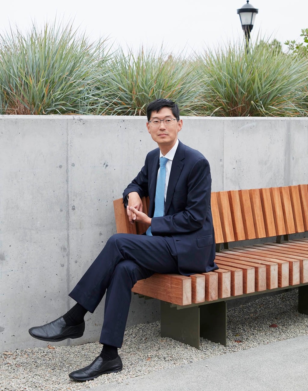
Stanford’s outstanding fundamental sciences research and advanced core technologies have helped fuel progress in the field, said Frank Longo, MD, PhD, the chair of neurology and neurological sciences and the George E. and Lucy Becker Professor of Medicine.
“We have strong basic science, a deep culture of interdisciplinary collaborations and the availability of resources, like great imaging capabilities, that allow us to do experiments more efficiently,” Longo said.
Here are just a few of the many projects in which Stanford Medicine scientists are restoring abilities that are crucial for patients’ daily living — and in some cases striving to prevent their loss in the first place.
Overcoming cognitive loss
Taking a different
approach on Alzheimer’s
Much of the effort to treat Alzheimer’s disease has focused on the protein known as amyloid, which forms sticky plaques that clog the brain and contribute to neurodegeneration.
But Longo has taken a different tack.
“I think that with Alzheimer’s and some of these other degenerative diseases, there are multiple forces that promote degeneration,” with amyloid being just one of them, he said. “We wanted to create a therapy that could address multiple mechanisms at one time.”
Normally, neurons respond to signals to maintain or shut down their synaptic connections, an essential part of the brain’s communication system. Some of these connections are lost naturally as we age, but in Alzheimer’s, the signal to kill these connections becomes overly active, Longo said. That leads to memory loss and other cognitive impairments.
Early in their Alzheimer’s research, Longo and his colleagues zeroed in on a molecule on the surface of neurons that regulates the network signals involved in this degenerative process. They then developed a synthetic molecule that binds to it to block the destructive process and promote regeneration. That molecule was C-31.
“We wanted to create a therapy that could address multiple mechanisms at one time.”
Frank Longo, MD, PhD, the chair of neurology and neurological sciences
In studies with mice, they found that C-31 made the neurons resistant to the effects of amyloid, prevented the formation of the toxic tau proteins that occur in the brain in the later stages of Alzheimer’s, decreased inflammation and reversed some of the effects of aging on the cells, like the shrinkage of neurons, he said.
“Our hope was that doing all of these four things might have a more powerful effect than just removing amyloid,” Longo said.
“One of the great mysteries in our field now is that we see people — even at advanced ages — with a brain full of amyloid but with memory and other cognitive function intact. While we do not understand this phenomenon and why it occurs in only a minority of people, we think we have created a compound that confers a therapeutic version of amyloid resilience.”
In mouse studies, the compound not only prevented damage to the synapses but also restored one of their most delicate structures — the dendritic spine, a protrusion in nerve cells that helps them communicate.
“We can apply it in a late state in the disease, when the dendritic spine is lost. The animal recovers to the levels of a young mouse,” he said. “It’s truly a regenerative effect.”
The beauty of the compound is that it can cross the blood-brain barrier, so it can be taken in pill form, making it easy and inexpensive to administer, Longo said.
Researchers in Europe recently completed a clinical trial to evaluate the molecule’s safety and explore ways to measure reductions in brain degeneration.
The trial included 242 patients with mild or moderate Alzheimer’s disease and was conducted by a company that Longo founded. Analysis of the trial is underway and, if indications are positive, the next step is a much larger trial to test for efficacy.
Longo and his research team are exploring how this experimental drug works and are finding additional conditions, such as Parkinson’s and Huntington’s disease, for which it might be useful. They have also found that C-31 may be able to counter nerve damage caused by the common cancer chemotherapy drug cisplatin.
“It gives us another entry point to better understand the mechanisms underlying these diseases, and in an exciting way, to gain insight into the emerging topic of brain resilience. This knowledge will help us develop additional, entirely new approaches,” he said.
More information on active Alzheimer’s disease trials available at Stanford is on the website of the Iqbal Farrukh and Asad Jamal Alzheimer’s Disease Research Center, which was renamed in 2021 in recognition of a donation made by the Good Planet Foundation: med.stanford.edu/adrc.html.
Looking for new
Alzheimer’s clues in genes
Neurologist Michael Greicius, MD, began his latest quest for a new Alzheimer’s drug in 2014, when he met a 57-year-old woman in the throes of advanced disease. She came to the clinic with her parents, both in their 70s.
Genetic tests showed that the patient had one copy of the gene for APOE4, a protein involved in cholesterol metabolism that is also thought to affect brain function.
People with the gene are at greater risk of Alzheimer’s, but those with two copies carry a risk that is extremely high. Remarkably, the patient’s mother had two copies of the APOE4 gene, yet she was in excellent health.
“That is when I scratched my head,” said Greicius, the Iqbal Farrukh and Asad Jamal Professor. “She had a double risk. She’s perfectly healthy and yet her daughter, with only one APOE4 gene, is already affected. Something is protecting the mother. I pretty strongly suspect it’s a gene.”
He resolved to look for rare genetic variations that could be protective — something the mother had but her daughter hadn’t inherited.
“The idea would be to try to find drug targets in those molecular pathways — mimic what these people have in their natural genomes,” said Greicius, who directs the Stanford Center for Memory Disorders.
Greicius is four years into the NIH-funded study for which he and his colleagues have amassed a collection of sequenced genomes from more than 500 people with and without Alzheimer’s. About half of these people are “protected” APOE4 carriers like the patient’s mother.
“That is when I scratched my head. … Something is protecting the mother. I pretty strongly suspect it’s a gene.”
Michael Greicius, MD, a professor of psychiatry and behavioral sciences
The researchers have obtained extensive biologic data for some participants through clinical exams, spinal taps, brain imaging, immunologic testing and skin biopsies. Greicius is screening the material from these individuals, looking for genes that might have a protective effect.
He’s also examining patients at the opposite end of the spectrum — those who don’t have the high-risk APOE4 gene but who develop Alzheimer’s at an earlier age, before they reach 65. This could point to previously unknown variants that could be implicated in the disease, he said.
Once these genes are identified, researchers can pinpoint the proteins they produce, then develop new drugs that may be able to block damaging proteins or enhance protective ones and, as a result, slow or stop the degenerative process.
Greicius’ work has already borne fruit. He analyzed 25 independent studies and showed that a common genetic variation known as Klotho-VS, which protects against age-related cognitive decline, reduces the risk of Alzheimer’s by 25% to 30% in older people who carry the risky APOE4 gene. He published papers on the work in JAMA Neurology in April 2020 and in Neurobiology of Aging in January 2021.
“That was a reassuring example that these variants are out there,” he said. “Thirty percent is good. We’re looking for variants that reduce risk by 80 or 90%. But this is certainly a good start.”
More information is on the website of the Iqbal Farrukh and Asad Jamal Alzheimer’s Disease Research Center: med.stanford.edu/adrc.html.
Fighting cognitive decline
by taming inflammation
Scientists have long focused on inflammation as a major cause of cognitive decline among patients with Alzheimer’s and other neurodegenerative diseases. But they have never understood the mechanisms behind it.
Katrin Andreasson, MD, a professor of neurology and neurological sciences, recently identified a possible pathway for inflammation in the brain and found a way to inhibit it to restore cognitive function, a finding she described in a January article in the journal Nature.
The key, she found, lies with a group of immune cells known as myeloid cells, which are among the body’s first line of defenders. In the brain, myeloid cells are known as microglia, which also help clean up debris (like the plaques in brains of people with Alzheimer’s) and control inflammation levels. In the blood, these cells are the macrophages and monocytes.
In her experiments, Andreasson compared these immune cells from older people (over 65) with those from younger people (under 35) and found the cells change dramatically as we age. In older brains, microglia promote a damaging, hyper-inflammatory environment instead of maintaining calm.
Andreasson’s research revealed a downward spiral of events that begins with older cells producing significantly more of the hormone prostaglandin E2, which regulates inflammation in the body.
She detailed other molecular changes in which more of the hormone molecules bind to cells, ultimately depleting the cells’ energy stores and leaving them in a perpetually exhausted state. The cells essentially devolve from young to old. Surprisingly, the changes occur not only in the immune cells in the brain but also in the macrophages in the blood, she said.
Most importantly, Andreasson and her colleagues tested older and younger lab mice using two compounds known to block the binding of the hormone and the molecule it attaches to on the cell — the EP2 receptor. They were able to stop the damage from occurring in the cells in the brain, as well as in the blood.
“If we could somehow change our microglia so they are behaving in a healthier way, that might go a long way toward slowing down the process of Alzheimer’s disease.”
Katrin Andreasson, MD, a professor of neurology and neurological sciences
“We were able to restore cognition to a youthful level,” she said, as the older mice were able to navigate a maze just as well as young ones. “What was a real shock was when we tested it in the circulating blood (outside the brain). … We found if you block an EP2 receptor in a macrophage, you could restore youthful metabolism.”
That means it may be possible to devise a drug that preserves cognitive function but doesn’t have to reach the brain. “That’s good news,” she said, “because every time you put something into the brain, there is potential for side effects.”
Scientists haven’t tested either compound in humans, so the drugs’ toxicities aren’t known, Andreasson said. But it’s a promising avenue for scientists to pursue in preventing cognitive decline.
“If we could somehow change our microglia so they are behaving in a healthier way, that might go a long way toward slowing down the process of Alzheimer’s disease,” she said.
More information about Alzheimer’s disease research and treatment is on the National Institutes of Health website: nia.nih.gov/health/alzheimers.
Getting moving again
Restoring hand function
through nerve transfer
People who have lost use of a hand because of spinal cord injuries or stroke now have an option for regaining movement: It’s called nerve transfer, a microsurgical technique that has emerged in the past five to 10 years, said Thomas J. Wilson, MD, clinical associate professor of neurosurgery.
In nerve transfer surgery, surgeons steal a functioning nerve with a less critical role and stitch it to a damaged nerve. The functioning nerve then regenerates through the damaged nerve to reestablish nerve supply to the target muscles and restore function. It can take as long as two years for patients to regain movement because the nerve grows very slowly and has to work its way into the muscle, he said.
“It’s a rob-Peter-to-pay-Paul phenomenon,” Wilson said. “You can steal something less important and give it to a more important movement.”
He has had good results using this technique in patients with spinal cord injuries. These patients report valuing hand function even more than walking, he said, because use of their hand increases their independence, allowing them to feed and dress themselves and to manually operate a wheelchair.
For the past year, Wilson has participated in a national clinical trial, sponsored by the U.S. Department of Defense, to track 70 spinal cord injury patients who are undergoing nerve transfer. The goal is to better predict which patients will do well after the surgery and to characterize the results they experience.
Wilson is also among a handful of neurosurgeons in the country using nerve transfer surgery to restore arm use in stroke patients. This procedure is more complex because the disabled limb is not a useful source of functioning nerves: The original injury is in the brain and broadly impacts nerves in that limb.
“Traditionally, nerve transfer surgery has been used for nerve injuries, but we are starting to think outside of the box, and we are applying this technique to other patients, including patients with spinal cord injury, stroke and traumatic brain injury.”
Thomas J. Wilson, MD, a clinical associate professor of neurosurgery
Instead, he swipes a nerve from the opposite limb. For instance, in someone with left-sided weakness, he cuts a segment of the cervical 7 (C7) nerve on the right side of the body, brings the nerve across the neck, and connects the right C7 nerve to the left C7 nerve to restore left-arm function. The C7 nerve’s function overlaps with that of other nerves, so it can be sacrificed in the unaffected limb without significantly compromising its use.
Normally, the body’s left side is controlled by the right side of the brain, and vice versa. But in this case, the hope is that the brain will adapt to allow the left side to assume control over the left limb.
“It turns out it actually does work,” Wilson said. A functional MRI, which maps the area of the brain being activated, shows that after a successful nerve transfer to the left side, the left region of the brain lights up, he said.
Wilson has treated three stroke patients with the surgery, two of whom are far enough into recovery to show improvement in arm function. These patients are able to dress, bathe and feed themselves using their once-paralyzed limb.
He said other neurosurgeons have used nerve transfer surgery in people with traumatic brain injuries, as well as those with cerebral palsy, to restore or improve hand and arm function.
“Traditionally, nerve transfer surgery has been used for nerve injuries, but we are starting to think outside of the box, and we are applying this technique to other patients, including patients with spinal cord injury, stroke and traumatic brain injury. The results have been very promising, but there is still a lot to learn in order to optimize our patient selection and outcomes,” he said.
“The next major hurdle is re-educating the medical community and making them aware that these techniques are available. I think there are probably a lot more people we could help if more clinicians were aware of what we have to offer.”
More about the procedure is on the Stanford Center for Peripheral Nerve Surgery website: stan.md/nervesurgery.
Improving brain implants
to treat Parkinson’s disease
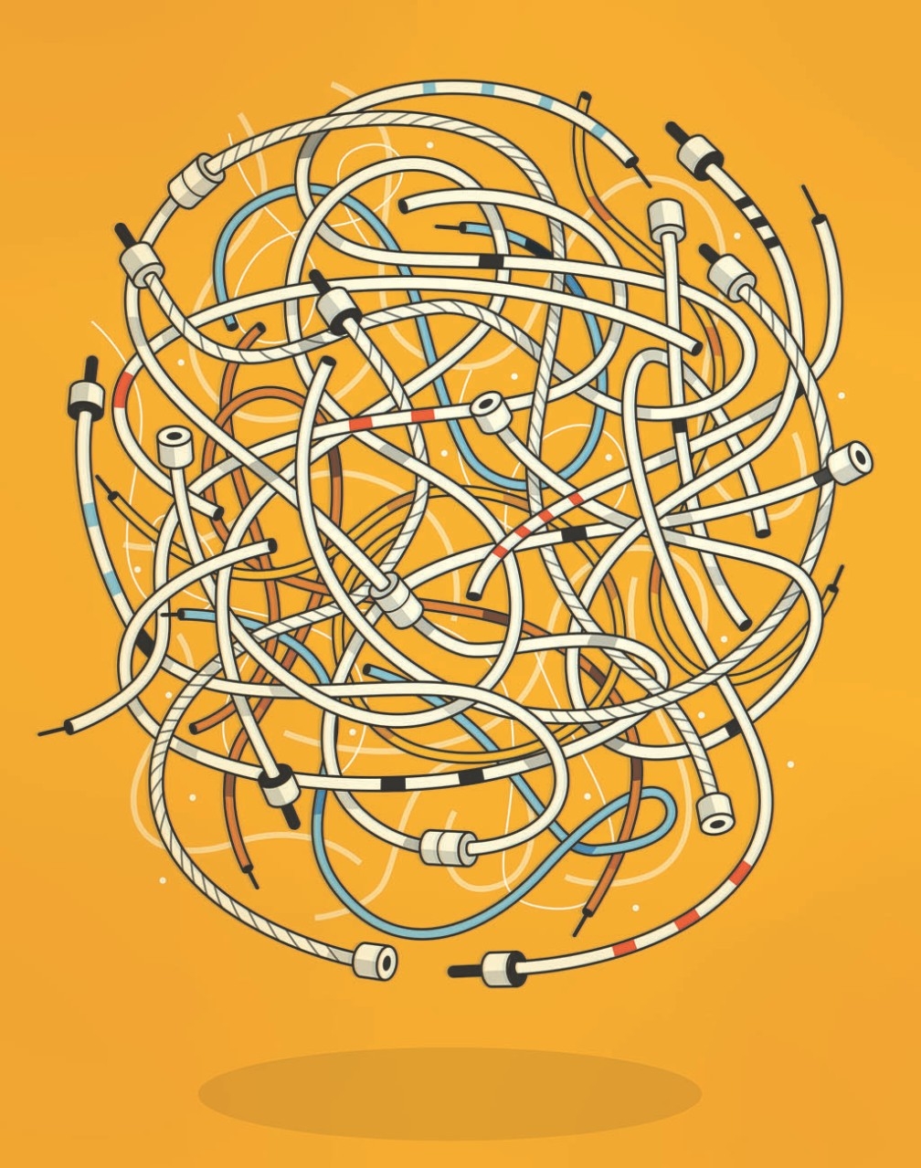
The brain may look like a big scoop of spaghetti. But it’s really an immensely complex electrical device whose component nerve cells, or neurons, are analogous to insulated, current-carrying wires.
Helen Bronte-Stewart, MD, the John E. Cahill Family Professor in the department of neurology and neurological sciences and chief of that department’s movement disorders division, is spearheading an effort to boost the ability of electrodes implanted in the brain to treat Parkinson’s disease.
Parkinson’s, the second most common neurodegenerative disease, affects 10 million people worldwide, according to Bronte-Stewart, the director of the Stanford Comprehensive Movement Disorders Center.
The motor-impairment aspect of the disease stems from the mysterious die-off of a set of neurons in the midbrain that form part of the sensorimotor network. One of the consequences of the die-off is that neurons in this network acquire an overly pronounced tendency to fire in sync at specific frequencies, akin to a brain arrhythmia. They begin transmitting prolonged spontaneous rhythmic bursts of movement-impairing signals instead of movement-shaping ones.
Medications can mitigate Parkinson’s symptoms — including visible tremor, faulty gait, limb rigidity, difficulty in initiating movements, slurred speech and, sometimes, impaired cognition. But they can also cause side effects and, as the disease worsens, fail to control symptoms, Bronte-Stewart said.
When medications fail, patients can benefit from an increasingly popular treatment called deep brain stimulation, or DBS, which restores control by disrupting the brain’s unwanted rhythmic firing.
Approved in 1997 for Parkinson’s disease, deep brain stimulation involves embedding electrical leads in the brain (most often the subthalamic nucleus) to act as a kind of anti-noise system.
Driven by a battery-operated pulse generator implanted in the chest, the leads fire their own trains of electrical pulses in the appropriate spot, countering the errant outbursts that cause Parkinson’s symptoms.
With standard DBS, the stimulator-driven pulse train flows steadily, changing only when the physician adjusts the patterns, on a trial-and-error basis, to maximize tremor inhibition and gait improvement without triggering side effects such as slurred speech, sensory disturbances, involuntary muscle contractions or balance problems.
In 2013, the FDA approved, for experimental purposes, a version of the implanted pulse generator that not only sends electrical bursts to the brain but also can record how the brain neurons are firing.
Researchers could now accumulate data on brain-signaling patterns in the vicinity of the implanted electrodes while the patient was walking, speaking, sitting, sleeping or engaging in other activities.
In June 2020, the FDA approved the commercial implantation of this “listening” device, making it much easier for physicians to make therapeutically useful setting adjustments because they can read brain signals from the device instead of inferring them from a patient’s motion, posture and comments.
Bronte-Stewart intends to further optimize and personalize this feedback. She is the principal investigator on a global trial of an advanced version of DBS called adaptive DBS. The goal is to transmute the accumulated data of years of research into an algorithm that lets the pulse generator do the reading in real time and, in response to what the brain is doing, directly alter its signaling pattern.
DBS was first approved in 1991 for essential tremor, a movement disorder that’s more common than Parkinson’s disease. It’s also approved for some types of dystonia, a movement disorder in which a person’s muscles contract uncontrollably; for epilepsy; and, in certain cases, for obsessive-compulsive disorder.
DBS is also being tested in a clinical trial led by Jaimie Henderson, MD, professor of neurosurgery, to treat reduced consciousness induced by brain trauma.
DBS device implantations have been performed on about 200,000 patients worldwide, close to 1,500 of them at Stanford.
For more information on deep brain stimulation to treat Parkinson’s disease, see stan.md/DBS.
Restoring movement for stroke
patients through stem cell transplant
Neurosurgeries with stem cells have demonstrated just how resilient and adaptable the brain can be. In multiple studies, Gary Steinberg, MD, PhD, has used stem cells in stroke and traumatic brain injury patients to restore their ability to walk, speak and return to some of their normal activities.
Steinberg published results from a landmark trial in 2016 in the journal Stroke in which he injected bone marrow-derived stem cells into an injured area of the brains of 18 patients.
Three-quarters of the patients had clinically meaningful recoveries, meaning their daily lives were changed for the better. The others had slightly less improvement or remained the same. The recovery of some of the patients was dramatic — they were able to run and speak again after having been trapped in their injured bodies.
“Those circuits that we thought were dead in stroke patients were not irreversibly damaged,” said Steinberg, the Bernard and Ronni Lacroute-William Randolph Hearst Professor in Neurosurgery and Neurosciences. “They were repressed and could be resurrected.”
Steinberg has since been examining the underlying mechanisms of these recoveries. In MRI images of patients taken after the procedures, he observed a transient signal near the injured area — a bright spot — that correlated with how well the patients fared over the longer term. He speculated that this signal might indicate a beneficial inflammatory response, which his recent lab studies have borne out.
He found that the stem cells were not creating new neurons, as he initially thought, but were releasing dozens, if not hundreds, of different healing molecules. These molecules include growth factors that build new nerve fibers and proteins that help create blood vessels, as well as a number of immune system cells that can enhance brain repair.
“Those circuits that we thought were dead in stroke patients were not irreversibly damaged. They were repressed and could be resurrected.”
Gary Steinberg, MD, PhD, a professor in neurosurgery and neurosciences
“It turns out that the beneficial inflammatory response is present not just where the lesion is but is more widespread throughout the brain,” he said. “It probably stimulates circuits very widely throughout the brain.”
Steinberg has tested the same stem cells as part of a multi-center trial involving patients who suffered traumatic brain injuries at least a year before the treatment. As in the stroke study, after six months, the treated patients showed significant improvement in their ability to move and walk, compared with control patients. The researchers reported the results in the journal Neurology in January 2021. The most common side effect was headaches, likely related to the surgical procedure, the scientists reported.
Steinberg is embarking on a study of a different kind of stem cell — neural stem cells derived from human embryonic tissue, known as NR1 cells. These stem cells, which he developed 20 years ago, have advantages: They are easier to grow than bone marrow-derived cells, can be manufactured in large quantities and are not genetically altered.
He plans to begin testing them this year in a Stanford-sponsored, first human trial in about 20 chronic stroke patients with partial paralysis. The procedure involves transplanting the cells directly into the brain near the area of the injury. Steinberg is the only investigator in North America using direct brain transplantation of stem cells for stroke.
“We expect that if this strategy works, we will be extending it to other indications like traumatic brain injury, spinal cord injury and, hopefully, even neurodegenerative diseases like Parkinson’s, ALS or, ultimately, Alzheimer’s, though that’s quite a bit in the future,” he said.
For more information on participating in the trial, email stemcellstudy@stanford.edu.
A high-tech glove could enable
stroke patients to rehab at home
Another new approach to treating patients who’ve suffered strokes could come from the wearable technology field.
By 2030, nearly 4% of American adults will have had a stroke, according to the American Heart Association, and as many as 80% of those who survive will end up with weakness and loss of sensation in their arms and hands.
“Having the use of two hands is absolutely essential for normal functioning. But currently there aren’t many effective interventions that can help people get that function back following a stroke,” said Caitlyn Seim, PhD, a research fellow at the Wu Tsai Neurosciences Institute at Stanford.
Most health insurers cover a limited amount of exercise-based stroke rehabilitation, and half of stroke survivors don’t have the mobility to even access these programs. To close this gap, Seim engineered a high-tech glove that she and her collaborators hope will one day let stroke survivors recover lost function in the comfort of their homes.
The gloves use haptic technology — originally developed for the video game industry to simulate interacting with objects and other sensory experiences — to stimulate patients’ hands with programmed patterns of vibration.
Researchers have hypothesized that applying vibration to specific muscle and sensory receptors in the hands could trigger a long-term rewiring of the brain, allowing people to regain control of their weakened limbs.
More immediately, the vibrations could also help relieve involuntary muscle contractions which distort patients’ limbs and constrict movement.
“A vibrating glove that improves hand function after stroke would be a breakthrough in the field of stroke rehabilitation.”
Maarten Lansberg, MD, PhD, a professor of neurology
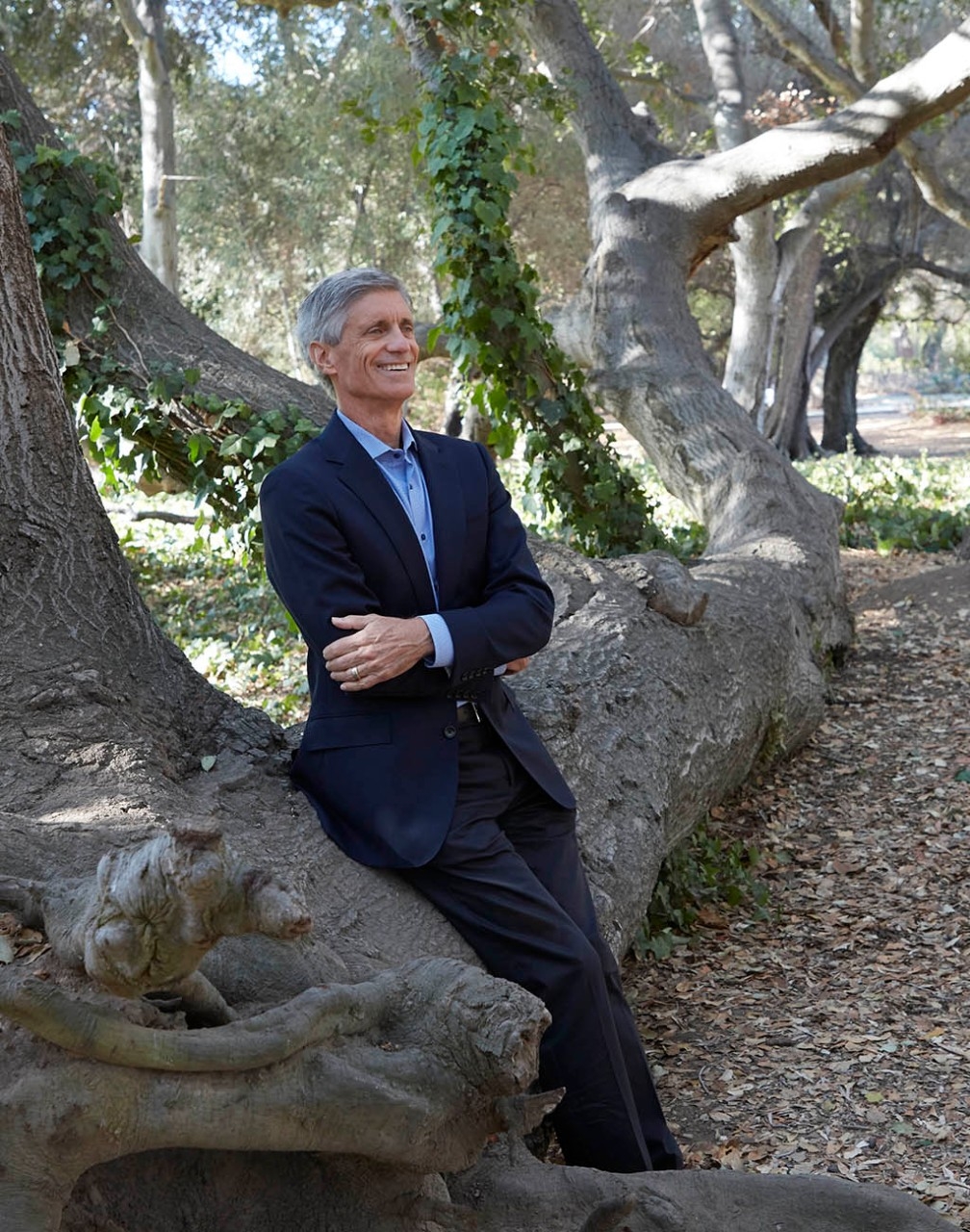
This idea has not been tested outside of limited laboratory studies, but that will change with Seim’s new wearable technology, which she is designing for real-world use in collaboration with Stanford Medicine stroke expert Maarten Lansberg, MD, PhD, a professor of neurology, and haptics expert Allison Okamura, PhD, a professor of mechanical engineering.
“A vibrating glove that improves hand function after stroke would be a breakthrough in the field of stroke rehabilitation,” said Lansberg. “Dr. Okamura and I are very excited about this technology, which can be easily used by people in almost any environment.”
The research team has designed the gloves to be easy to use in a home setting by patients who suffer a wide variety of stroke-related symptoms.
“Patients need to be able to put them on themselves and wear them comfortably at home, whether they have really tight fingers or really weak fingers,” said Seim, whose work is supported by grants from the Wu Tsai Neurosciences Institute and the National Center for Medical Rehabilitation Research.
The team has enrolled 20 patients in a clinical trial to test how well the gloves work in a home setting. Patients will use the gloves for two months, then researchers will monitor hand function for up to six months. A second trial is underway to determine how haptic stimulation affects communication between hand and brain.
“So far, everyone who’s finished with the device says they miss it, they want it back, they love it,” Seim said. “And this is after we made them wear the glove for 160 hours. So I think that’s a promising sign.”
Lansberg and neurology and neurosurgery professor Marion Buckwalter, MD, PhD, who direct the Stanford Stroke Recovery Program, are also adapting gaming technology to help patients recover hand function. A study published in March in the rehab-focused journal PM&R found that patients who used a virtual reality rehabilitation gaming device for eight weeks at home showed marked improvement of hand function and were highly satisfied with the device. The team is testing this approach in a larger, randomized controlled clinical trial.
More about efforts to improve mobility and other functions after stroke is on the Stanford Stroke Recovery Program website at stan.md/strokerehab.
New ways to see
Restoring sight to the blind
with a retinal implant
After more than 15 years of research, Daniel Palanker, PhD, and his collaborators have produced and successfully tested a first-generation retinal implant that can restore vision in people with age-related macular degeneration.
The eye disease leads to a gradual loss of sight in the center of the visual field because of damage to light-sensing nerve cells in the retina, called photoreceptors. Palanker’s lab has developed a technology that does the job of photoreceptors — a photovoltaic implant that converts incident light into electric current and transmits the visual information to the remaining, intact inner retinal cells.
“We are just replacing one layer of cells that has been lost with photovoltaic pixels,” said Palanker, a professor of ophthalmology. “We use the rest of the retina to process the electronic visual input and thereby help restore sight.”

A company that has licensed his technology from Stanford tested the first generation of the device (called PRIMA) with 100-micron pixels in five patients in France. Four of them achieved visual acuity close to the 20/420 limit set by this pixel size, he said. With electronic zoom, they were able to read letters four times smaller (20/100) on a vision chart.
Moreover, they could simultaneously use the prosthetic for central vision along with their remaining natural peripheral vision. Palanker and his colleagues published the findings in March 2020 in the journal Ophthalmology.
“It’s a very exciting confirmation of many assumptions we have made at the beginning of a very long journey,” Palanker said. Researchers will now begin a larger clinical trial of the implant in 38 patients in Europe and in the United States, including Stanford.
Macular degeneration is the most common cause of untreatable blindness in the United States among people 50 and older. Drug injections in the eye can minimize vision loss in some forms of the disease, but it goes only so far in preventing blindness.
“It’s a very exciting confirmation of many assumptions we have made at the beginning of a very long journey.”
Daniel Palanker, PhD, a professor of ophthalmology
Palanker’s device consists of a 2-millimeter chip that is surgically implanted under the retina. The procedure takes about two hours, often under general anesthesia to minimize a patient’s movement. With the chip in place, patients don augmented-reality glasses with a small video camera on the rim.
The camera captures images, and the glasses project them onto the chip implanted under the retina using invisible near-infrared light. Each pixel in the chip converts the incoming light energy into an electric current, much the way a solar panel converts sunlight to electricity, Palanker said.
The electric current flowing through the tissue stimulates the nearby neurons, which relay these signals to the rest of the retina and ultimately to the brain, which decodes the image so the patient can see.
In a recent preclinical study, submitted to a Nature Portfolio journal, Palanker’s group demonstrated much higher resolution in rodents by making the pixels as small as 20 microns. If these implants work well in human patients, they could achieve 20/80 vision; with double magnification, they could see well enough to drive, he said.
“I think these implants will be affordable because the fabrication technology is scalable to large numbers, as with any silicon chip,” he said.
Palanker is a consultant for the company that licensed the technology and an inventor of the Stanford-licensed patents.
For more information on the retinal implant, see stan.md/retinaimplant.
Building an artificial retina
Neuroscientist E.J. Chichilnisky, PhD, is also developing a device to help restore vision for people with retinal disease, but his approach is different. His group is designing an artificial retina — an electronic implant that replicates the complex process by which key nerve cells, known as the retinal ganglion cells, convey visual information to the brain.
The advantage of this approach, compared with Palanker’s device, is that it bypasses the photoreceptors and targets the underlying retinal cells that have a direct communication line to the brain.
There are more than a million of these cells in the inner layer of the retina but, unlike photoreceptors, they are not uniform.
There are some 20 types of retinal ganglion cells, each with a different role in conveying visual stimuli to specific areas of the brain. The researchers have to learn the language of each of these cells and how each communicates with the brain.
“What we are doing is developing a smart device that records the activities of these cells, uses that information to figure out who is who and how to target each of these cells individually with customized information so they can send the right signals to the brain,” said Chichilnisky, the John R. Adler Professor of neurosurgery and of ophthalmology. “It’s a high-end kind of interface.”
In other words, the scientists have to faithfully reproduce the way the cells encode visual stimuli so the brain responds with an accurate visual image.
“We do want to restore vision to the blind, but we also believe this technology could have implications for other areas of the brain while producing a spectacular scientific instrument to understand the visual pathways.”
E.J. Chichilnisky, PhD, a professor of neurosurgery and of ophthalmology
The device could help the millions of patients who have macular degeneration or retinitis pigmentosa, conditions caused by lost or damaged photoreceptor cells. The retinal implant would bypass these cells to restore vision.
As part of the Stanford Artificial Retina Project, Chichilnisky is collaborating with Palanker and about 20 other scientists, including experts in electrical engineering, retinal surgery, neurophysiology, computational neuroscience and visual behavior. The project kicked off six years ago, when the group received a Big Ideas in Neuroscience grant from the Wu Tsai Neurosciences Institute.
The researchers have developed a prototype chip and a series of advanced algorithms that they have been testing in animal models and in donated human retinas. They are refining the technology and hope to have a 2-millimeter implant in two to three years so they can begin human trials.
“We do want to restore vision to the blind, but we also believe this technology could have implications for other areas of the brain while producing a spectacular scientific instrument to understand the visual pathways,” Chichilnisky said.
In the near term, he said, the research could benefit patients such as those with Parkinson’s disease by improving techniques for deep brain stimulation. Surgeons implant an electrode that activates neurons in a specific area of the brain, but how the method works is not well understood.
The retinal project could shed light on the natural patterns of the underlying brain circuits and how to interface with them, and thus help make deep brain stimulation more targeted and effective. It also might contribute to other brain interfaces to help people with memory loss, paralysis or other disorders tied to the brain.
For more details on the Stanford Artificial Retina Project, go to: artificial-retina.stanford.edu.
Using drugs to tackle
glaucoma at its source
Ophthalmologists typically manage glaucoma — the world’s leading cause of blindness — through various methods to lower the fluid pressure within the eye. Over time, elevated pressure damages retinal ganglion cells and their long projections, known as axons, that form the optic nerve. This degenerative process kills the optic nerve and results in blindness.
Jeffrey Goldberg, MD, PhD, professor and chair of ophthalmology, believes it will not be enough to focus exclusively on managing internal eye pressure. Rather, he sees the future of glaucoma care in new therapies that preserve and protect the retinal ganglion cells and their axons and, possibly, regenerate those that are lost.
“For many years, it was thought that vision restoration trials weren’t possible — that you needed too many patients, that the disease was slow and variable so it would take too many years and be too expensive,” said Goldberg, the Blumenkranz Smead Professor and director of the Spencer Center for Vision Research.
But his recent experience has shown it is possible to conduct short-term trials and generate encouraging results.
One study involves a molecule called C1q. This molecule’s importance in pruning nerve-cell connections was discovered by the late Ben Barres, MD, PhD, former chair of neurobiology. C1q is believed to underlie many neurodegenerative processes, including the destruction of retinal ganglion cells.
“What we’ve learned through all these trials is that we can do them in a reasonable fashion and time frame and start to address this big unmet need of vision loss in glaucoma.”
Jeffrey Goldberg, MD, PhD, a professor and chair of ophthalmology
A South San Francisco, California, company co-founded by Barres has developed a monoclonal antibody that binds to C1q and inhibits its activity. In a Phase 1 trial, Goldberg and his colleagues tested the antibody in the first human trial by injecting it into the eyes of glaucoma patients.
“We did a molecular characterization and showed that the antibody drug was indeed mopping up all the free C1q from inside the eye,” he said. The next step is to see if it can improve vision.
In other trials, Goldberg and his colleagues have tested different nerve growth factors that nourish and maintain nerve cells. In one study, they have experimented with an implant filled with cells genetically engineered to make ciliary neurotrophic factor, a naturally occurring molecule shown in animal studies to protect and regenerate axons in the optic nerve.
The scientists implanted a 1-by-5-millimeter capsule into the middle of the eye, where it released a steady flow of the growth factor onto the retina and optic nerve. The results showed a thickening of the nerve fibers, which is encouraging, as these fibers typically thin out as glaucoma progresses, Goldberg said. The scientists are testing the use of two implants, instead of one, and treated their first two patients this spring and early summer.
“The implants look great, but it will take some time to measure their effects,” Goldberg said.
In a separate trial, researchers tested eyedrops containing high doses of human nerve growth factor, a naturally occurring protein that similarly supports nerve cells. They enrolled 60 glaucoma patients at Stanford over the course of a few months in a randomized trial designed to gauge safety. These results were also encouraging, with some signs of nerve fiber thickening and a great safety profile.
“The ideal next step is to test the eyedrops for a full year to see if they help improve the patients’ vision,” Goldberg said.
Whether these or other candidate therapies in clinical trials for glaucoma could prove effective, pushing the field to complete such trials is showing a positive effect, he said.
“What we’ve learned through all these trials is that we can do them in a reasonable fashion and time frame and start to address this big unmet need of vision loss in glaucoma.”
For more on these trials, visit: stan.md/glaucomadrops.
Bruce Goldman and Nicholas Weiler contributed to this article.
— Contact the authors at medmag@stanford.edu
