


Cartilage can’t heal well — but that may change
New scaffolds, drugs and imaging aim to repair joints and prevent arthritis
Running, jumping, throwing or typing. All involve the vigorous movement of muscles, tendons and bone through joints. But there’s a hidden player: articular cartilage. This tissue that lines joints is one of the slipperiest surfaces around, and cartilage on cartilage generates less friction than ice on ice.
Without it, joints grind and bone wears away or deforms, making simple movement — like stepping up stairs or raising an arm to reach a glass on a high shelf — painful.
But cartilage is relatively inert. No blood vessels provide nutrients, no true stem cells have been definitively identified and few immune cells patrol healthy joints. As a result, cartilage has little capacity to regrow once it’s been lost. This leaves tens of millions of Americans struggling with chronic joint pain (largely due to arthritis), limited mobility and no good treatments.
Related content
- Lessons from antlers: How 3D-printed scaffolds and antler stem cells advance bone repair
Stanford Medicine researchers are tackling cartilage loss, from spurring cartilage cells into new cycles of regeneration to designing body-friendly scaffolds to guide their regrowth. They’re also devising better imaging techniques to identify the earliest stages of cartilage damage and ways to test interventions on a person’s own cells grown in a lab.
Looming large, however, are many clinics promoting unproven treatments, including injecting a person’s homogenized fat cells or blood plasma into their affected joints. In the absence of effective treatments, desperate people pour money and hope into pipe dreams peddled by would-be miracle workers with few qualifications.
Here’s a look at how researchers are attempting the seemingly impossible — regenerating or repairing cartilage that’s worn away, damaged or gone entirely.
Catching cartilage damage before it’s too late
Identifying invisible damage through powerful imaging and biomarkers
Constance Chu, MD, the Elsbach-Richards Professor in Surgery and vice chair of research in the Department of Orthopaedic Surgery, has spent more than three decades restoring anterior cruciate ligaments, menisci and cartilage surfaces while growing joint cartilage in the lab.
Her patients have ranged from teenagers whose ACL injuries put their seemingly healthy knee cartilage at risk to adults with no cartilage remaining who require joint replacement. Witnessing this progression from vulnerability to pain and degeneration has led Chu to focus her research and clinical practice on preserving joints.
The problem, she believes, is diagnostic. “Cartilage doesn’t have any nerves,” she explained, “so when you damage or injure cartilage, unless it’s a significant injury connected with injury to other tissues, you don’t even know about it.” Standard imaging — largely relying on joint narrowing visible on X-rays — detects only end-stage disease that is already too advanced to reverse.
Chu’s lab develops and evaluates MRI techniques that detect what she calls “invisible cartilage damage” — structural changes that might still be reversible but are invisible to conventional scans. “I call it giving injured and stressed cartilage a voice — or the ability to be seen if not heard,” she said.
“II would rather save the joint than replace it.”
Stuart Goodman, MD, PhD, the Robert L. and Mary Ellenburg Professor in Surgery
She has been particularly focused on patients who have suffered ACL tears — a typically young population whose cartilage she finds to be abnormally soft just weeks after injury. Her research shows signs of deterioration long before any symptoms appear. Specialized MRI scans performed one and two years after an ACL reconstruction detect cartilage changes in nearly half of those patients, her research has found; caught at that stage, those changes might be treatable. Left undetected, they likely progress to osteoarthritis just 10 to 15 years later, while most are in their prime work and child-rearing years.
Nidhi Bhutani, PhD, an associate professor of orthopaedic surgery, is trying to close the same diagnostic gap by identifying biomarkers in blood and joint fluid that yield a score of joint health. In collaboration with Chu, she has shown that the immune signatures of osteoarthritis can arise in patients with little to no imaging clues.
The findings suggest that osteoarthritis, historically treated as a uniform disease, encompasses distinct forms that genomics could help identify and map. “If you’re using the same drug for patients with different subsets of disease, of course the clinical trial fails,” Bhutani said.
The goal is to bring to orthopaedics the same precision that has transformed oncology: Matching the patient to the treatment, rather than averaging the results among everyone in the room.
Engineering scaffolds that regrow bone and cartilage
Microribbons seeded with regeneration-spurring molecules serve as cellular highways into damaged joints
Stuart Goodman, MD, PhD, the Robert L. and Mary Ellenburg Professor in Surgery, has been operating on joints at Stanford Medicine for over 40 years, focusing on rebuilding those destroyed by infection, failed prior surgeries or injuries that never healed properly. But he became increasingly aware of the limits of the technique.
“Putting in more plastic and metal in someone’s joint or around their joint, you run out of bone and cartilage and other tissues to work with,” he said. “I would rather save the joint than replace it.”
For Goodman, the frustration of running out of biological material to work with in the operating room sent him in a different direction: toward the question of what it would take to restore joints rather than replace them.
He spent years studying the literal fallout of joint replacement — the plastic particles shed by worn implants that trigger inflammatory cascades, eat away at surrounding bone and force ever more complex revision surgeries. “For 20 years, I focused on the byproducts of plastic and metals and how they’re not good for cells, and they destroy bone,” he said.
He gradually shifted his focus toward a larger ambition: not better artificial joints, but a biological approach that could make artificial joints unnecessary. The lowest-hanging fruit, he found, was bone. Bone’s relative capacity for repair — its blood supply, its marrow full of stem cells, its outer membrane that delivers blood and nutrients — makes it tractable in a way that cartilage is not.
What resisted solution was not small bone defects, but large gaps (around 2 centimeters in people) that cannot heal on their own. These gaps are often the result of severe trauma, infection or the removal of tumors. He built a lab to study how the immune system and the stem cells found in the marrow interact during bone healing, and how to modulate that interaction to promote repair.
“The opportunity is to intervene earlier, when it is starting, when we still have a chance to reverse it.”
Fan Yang, PhD, and director of the Stem Cells and Biomaterials Engineering Laboratory
Goodman has collaborated closely with Fan Yang, PhD, an associate professor of orthopaedic surgery and of bioengineering, and director of the Stem Cells and Biomaterials Engineering Laboratory. Yang studies why cartilage and large bone defects are so resistant to repair.
“Our body actually has all the necessary cells,” she explained. “The question is, how do we get the right ones to come at the right time and do the right things?”
Her answer was to design a scaffold that the body would accept and actively colonize — not a passive structural support, but a biological invitation. She developed what she calls
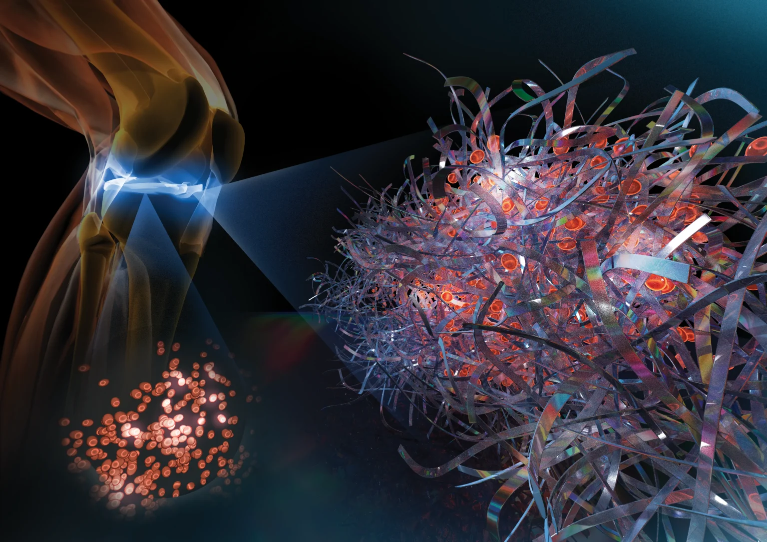
“microribbons” — flat, fettuccine-shaped hydrogel structures that interlock into a porous three-dimensional architecture.
Unlike conventional hydrogels, whose pores are too small for cells to migrate through freely, microribbons let cells move in quickly and begin producing their own tissue matrix. The ribbon architecture also gives the scaffold a springlike mechanical resilience — critical for cartilage, which must absorb and release compressive force with every step.
Yang has also focused on the immune environment surrounding the scaffold, developing surface coatings that shift local signaling from pro-inflammatory toward pro-regenerative — particularly important for bone, where immune cells from the marrow are constant participants in the healing process.
Goodman loads Yang’s microribbon scaffolds with engineered stem cells and tests them in animal models. The combination — an open, mechanically resilient scaffold delivering cells primed to tilt local macrophages from an inflammatory to a regenerative state — has produced promising results in both young and aged mice. But Yang’s most urgent message is also about timing.
“No regenerative therapy can treat those patients effectively once disease has reached end stage,” she said. “The opportunity is to intervene earlier, when it is starting, when we still have a chance to reverse it.”
How a ‘gerozyme’ inhibitor wakes up cartilage-making cells
Reversing aging by reprogramming existing cells
Cartilage, meanwhile, has resisted every structural approach that has worked for bone. With no blood supply, no known stem cells and no immune surveillance, there is extremely limited self-repair. Helen Blau, PhD, the Donald E. and Delia B. Baxter Foundation Professor and director of the Baxter Laboratory for Stem Cell Biology, has spent her career studying why some tissues regenerate and others don’t.
Blau and Bhutani teamed up to show that a fundamental assumption about cartilage — that regeneration would not occur without stem cells — was wrong. An experimental drug targeting an enzyme Blau calls a “gerozyme” can reprogram the damaged and aging cells already present in the joint, returning them to a healthier, more productive state. “It’s a reprogramming of the existing cells,” Blau explained. “It’s a completely new mechanism of regeneration.”
The gerozyme-blocking drug inhibits the age-related destruction of a molecule called prostaglandin E2, which Blau’s lab has shown to be essential to the function of muscle stem cells. They wondered whether the molecule might also play a role in aging cartilage and joints.
“It’s a reprogramming of the existing cells. It’s a completely new mechanism of regeneration.”
Helen Blau, PhD, director of the Baxter Laboratory for Stem Cell Biology
Bhutani and Blau found that the joints of old mice treated with the drug developed new, young-looking cartilage, and the animals exhibited less pain and greater function (as measured by gait, weight-bearing and sensitivity to touch before and after treatment).
Similar results were observed in animals with joint injuries like the ACL tears that frequently occur in people participating in sports such as soccer, basketball and skiing that require sudden pivoting. While the tears can be surgically repaired, about 50% of people develop osteoarthritis in the injured joint within about 15 years. A series of injections twice a week for four weeks of the gerozyme inhibitor after injury dramatically reduced the chance that the injured mice developed osteoarthritis.
Human cartilage samples obtained during joint replacement surgeries showed a similar pattern: Treated tissue produced more cartilage-building proteins than untreated controls and showed signs of cartilage regeneration.
“The fact that you can reprogram injured and diseased cells to cause regeneration is really like an anti-aging or a reverse-aging sort of effect — which I think is very, very promising,” Bhutani said. Blau and Bhutani are planning to bring the gerozyme therapy to clinical trials.
Precision medicine for building back cartilage
Patients’ in a dish, on a chip — sussing out
what works for you
Chu is also pioneering a precision medicine approach she calls joint avatars. These are miniature organoids — tiny three-dimensional mimics of human tissues or organs — grown from a patient’s blood or small tissue samples that allow her to estimate how that individual’s cartilage or joint lining tissue responds to different treatments before any intervention is undertaken.
She is collaborating with Bhutani and Yunzhi “Peter” Yang to create organoids specific to the patient and to the treatment being tested. The avatars offer a way to find out what will work for individual patients before subjecting them to it.
“You can potentially avoid having someone undergo an expensive, time-consuming procedure that isn’t going to work for them,” she said. “If that proves to be true, it’s very, very powerful.”
Goodman is pursuing a different route to the same problem: building the joint outside the body. His lab has designed a “joint-on-a-chip” system — a small plastic wafer engraved with fluid-filled channels that mimic the cellular composition and interactions that make up a functional human joint.
“We can take bone cells, cartilage cells, joint cells, immune cells and vascular cells, and connect them up in this microphysiological system,” Goodman said. “We can perturb it by adding, let’s say, a molecule that causes inflammation, and not only monitor it, but also use it to look at the effects of different drugs on that process.”
Where Chu’s avatars start from a single patient’s cells to predict how that person will respond to a treatment, Goodman’s chip is built to model disease itself — re-creating osteoarthritis, joint infection and inflammation in a controlled system where researchers can screen drug candidates long before they reach a clinical trial.
Developed with support from the National Institutes of Health’s national tissue chip program, in collaboration with the University of Pittsburgh, the project gives Stanford Medicine researchers a way to study how joint disease progresses and to test treatments without waiting on cartilage that, once damaged, still won’t regenerate on its own.
Fake treatments
Capitalizing on hope, unregulated clinics
promote unproven solutions
About one in four adults in the United States have been diagnosed with some type of arthritis, according to the Centers for Disease Control and Prevention, and about one in 10 report that arthritis or joint problems limit their daily activities.
The unproven treatment market — dominated by clinics offering injections of platelet-rich plasma, homogenized fat cells or mesenchymal stem cells into damaged joints — has grown large precisely because so many patients arrive at the end of their options.
The scientific premise behind mesenchymal stem cell injections isn’t baseless, and Stanford Medicine researchers are investigating their potential for joint repair. These stem cells are multipotent progenitor cells — found in bone marrow, fat and other tissues — capable of differentiating into cells called chondrocytes that build and maintain cartilage, among other cell types.
But that process is slow and exacting: In the lab, coaxing mesenchymal stem cells toward a cartilage fate typically takes weeks of culture with specific growth factors, delivered in a controlled, three-dimensional environment that mimics the joint’s biochemical and mechanical cues. Skip that step, and the cells have no particular reason to become cartilage at all.
The problem is that most clinics offering “stem cell” injections don’t even attempt to isolate or test these cells. They withdraw fat or bone marrow, spin it in a centrifuge and reinject the resulting mixture the same day. That mixture is a slurry in which true mesenchymal stem cells usually make up a tiny fraction of the cells, diluted among blood cells, immune cells and debris. But it has none of the purification, expansion or priming that legitimate research relies on.
“There’s really no biological basis for the treatments promoted by unregulated clinics working. If you see an effect, it’s placebo.”
Helen Blau
That gap — between what these specialized stem cells can plausibly do under carefully engineered conditions and what an unpurified, same-day injection can deliver — explains why these treatments so rarely outperform placebo in controlled trials. They are expensive, too. A same-day procedure can cost around $5,000 to $10,000.
Platelet-rich plasma, or PRP — falls into a similar gray area. Platelets are naturally occurring cell fragments in the blood that are packed with proteins that encourage cell growth and tissue repair. Although injection of PRP into damaged or arthritic joints has been shown to provide moderate pain relief and increased function for some people with mild to moderate knee arthritis, clinics marketing the “treatments” often claim much larger, universal benefits. The price at these clinics runs around $1,000 to $4,500 per course of treatment.
Chu, who led an NIH-funded conference on the responsible use of biologics in orthopaedic surgery, said this “one-injection-fits-all” approach ignores her PRP research finding that its effectiveness is highly variable and not well understood.
“Platelet-rich plasma is only as good as what you have to offer based on your age, your health, and your individual ability to repair and heal,” she said. In one study, she found that PRP from young, healthy people had an anti-inflammatory effect on cartilage tissue in culture, but PRP from older people with osteoarthritis actually stimulated inflammation, “which is not what you want.” Her development of joint avatars will allow her to identify which patients would benefit most.
Blau is direct about what the vacuum of available therapies for joint pain has been filled with. “People are desperate, so they go anywhere,” she said. “There’s really no biological basis for the treatments promoted by unregulated clinics working. If you see an effect, it’s placebo.”
What Stanford Medicine researchers are building — scaffolds that recruit the body’s own repair machinery, imaging sensitive enough to catch disease before it declares itself, organoids that let surgeons test what will work for a specific patient, and now a drug that reprograms the cells that were thought to be beyond reach — attempts to close that gap with something more durable.
