Lifting the burden of cancer
Advances in cancer science, prevention and care
Cancer is infamously cunning, expansive and relentless. It has a talent for evading treatment, spreading throughout the body and coming back again and again. Despite a steady decline in U.S. cancer mortality rates thanks to better screening and treatments, the absolute number of deaths from cancer continues to tick up, in part because of an aging population.
In response, scientists and clinicians are taking a page from cancer’s playbook, learning to be just as cunning, expansive and relentless in their efforts to beat back the disease. Cancer is meeting its match.
“The field of oncology has been completely transformed from where it was 15 years ago — many aspects of cancer treatment resemble science fiction now,” said Steven Artandi, MD, PhD, the Laurie Kraus Lacob Director of the Stanford Cancer Institute.
Index of topics in this article:
Stanford Medicine researchers are developing cellular therapies that trick cancer cells into self-destructing, and surgeons are using glowing dyes to sleuth out tiny tumors during surgery. They are inventing ever more sensitive ways to detect new or recurring disease before anything can be seen anatomically.
“When you can get information earlier, you can make precision health decisions,” said Joseph DeSimone, PhD, co-director of the Cancer Imaging and Early Detection Program at the Stanford Cancer Institute. “It lets you see the path emerging in front of you leading toward a disease state.”
His lab is developing 3D-printed microneedle patches that, when applied to the skin for just five minutes, can painlessly draw out enough lymphatic fluid for liquid biopsies. That could open up early detection to people who avoid blood draws because of cost or discomfort.
Increasingly, researchers are thinking creatively about every aspect of cancer care. They are looking beyond rogue cells and faulty genes to study how the gut microbiome can boost immunotherapy, how a patient’s mindset about cancer influences quality of life, and how tax credits for poor families enable behaviors that lower cancer risk.
Cancer cells continually evolve resistance to therapies, so researchers must relentlessly develop new treatments. Stanford Medicine has led the way in CAR-T therapy, which genetically programs a patient’s infection-fighting lymphocytes to home in on cancer instead. It’s approved by the Food and Drug Administration to treat lymphomas and leukemias as well as multiple myeloma. A new version, developed by Crystal Mackall, MD, the Ernest and Amelia Gallo Family Professor and professor of pediatrics and of medicine, is showing promising results in patients with relapsed B-cell lymphoma and is under expedited review by the FDA.
And last year, Stanford Medicine was the first in the world to treat a patient using a new cell therapy that takes advantage of lymphocytes with natural cancer-fighting abilities. Known as tumor-infiltrating lymphocyte, or TIL, therapy, it harvests lymphocytes that have infiltrated a tumor, multiplies them in the lab and infuses billions of them back into the patient.
The most original ideas are often born from unusual collaborations. In the 1950s, Henry Kaplan, MD, then a radiologist at Stanford Medicine, collaborated with nuclear physicists at SLAC National Accelerator Laboratory to develop the first medical linear accelerator in the Western Hemisphere and successfully treated the first patient, a 2-year-old boy with an eye tumor. Kaplan went on to become the founding chair of the Department of Radiology, which was later reorganized into several sections, including what is now the Department of Radiation Oncology, led by chair Quynh-Thu Le, MD. “We’re very proud of our legacy,” Le said.
The Department of Radiation Oncology’s diverse team of physicists, basic scientists and physicians work together to innovate and implement new drugs and radiation therapies for cancer patients. “We work hand in hand,” said Le, the Katharine Dexter McCormick and Stanley McCormick Memorial Professor II. “We cannot do one without the other.”
To foster more cross-pollination, the Stanford Cancer Institute was founded 20 years ago as a hub for research and clinical programs. “Stanford had been focused on cancer for decades, but it had been much more departmentally based, without a collaborative cancer center structure,” Artandi said.
Clinical trials a top priority
In 2016, the Institute was awarded Comprehensive Cancer Center status by the National Cancer Institute, a designation given only to cancer centers that excel in basic science research, translational research and clinical care.
“Clinical trials have been one of our highest priorities. Our strong belief is that research is standard of care in oncology,” said Artandi, the Jerome and Daisy Low Gilbert Professor.
This past year, about 1,000 patients enrolled in cancer clinical trials at Stanford Medicine, including a growing number of Phase 1 drug trials managed by the Early Drug Development Program. These first-in-human trials can be lifelines for patients who have run out of other options.
Stanford Medicine’s cancer centers saw nearly 250,000 clinic visits in 2024, which has doubled from about 10 years ago. Beyond the main center on the Stanford campus, the network includes centers in Los Gatos, Emeryville, Redwood City and Pleasanton. And a joint venture with Sutter Health is underway to build a state-of-the-art cancer center in Oakland that will expand Stanford Medicine’s reach to underserved communities.
Visits are expected to double again in the next 10 to 15 years, said Sridhar Seshadri, DBA, president of Stanford Medicine Cancer Center. “We want to leverage our significant strength in basic science and vibrant university ecosystem and move that toward the best cancer care for our patients,” Seshadri said.
Read on to learn about some of the work that’s inspiring a reimagining of cancer innovation and care at Stanford Medicine.
The Stanford Cancer Institute
Stanford Medicine’s hub for cancer research and care, the Stanford Cancer Institute, is recognized by the National Cancer Institute for prowess in stimulating novel laboratory, clinical and population-based cancer research and for finding ways to use these discoveries to improve patients’ lives. For more information visit med.stanford.edu/cancer.html.
LIGHTING UP TUMORS
Glowing dye helps surgeons find hidden cancer
George Poultsides, MD, hears the same question from nearly every patient he’s operated on: “Did you get all the cancer?”
As a young surgeon, he would confidently reply, “Yes.” Now, after seeing many patients with cancer that recurred, he tells them he removed everything he could see.
“It got me wondering if there was a way for us to enhance how surgeons see cancer,” said Poultsides, a professor of surgery who specializes in treating pancreas and liver cancer.
Many tumors are too small to identify or are hidden inside dense tissue, he noted.
Poultsides and his colleagues have experimented with fluorescent molecular dyes (in any color of their choosing — green stands out nicely) attached to antibodies that tumors absorb. They have found that the dye — visible under near-infrared light — allows them to safely locate and remove tumors.
The technique may be especially useful for laparoscopic surgery, during which surgeons are looking through scopes inside the abdominal cavity.
“Not finding all the tumors is the reason cancer surgeries fail,” Poultsides said. “If cancer returns a few years after an otherwise successful surgery, it is because of cancer cells that had escaped our notice.”
In a trial he led at Stanford Medicine, 11 patients with pancreatic cancer were intravenously infused with dye two to three days before surgery.
During the procedure, surgeons shone near-infrared light with a flashlight-like device into the abdominal cavity four times: after they first opened the abdomen; when they located the tumor; when they encountered lymph nodes in the abdomen, where metastases often appear; and after they had removed all the cancer they could see. Cancer foci as small as 2 millimeters in diameter glowed green under the light.
“The dye works,” Poultsides said. “It showed cancerous tissue that the radiologists and surgeons hadn’t seen,” though he added that they can’t know what they’re missing. The trial also showed that the dyes are safe, and it pinpointed an optimal dose.
Poultsides chose to run the trial in patients with pancreatic cancer because this cancer recurs in as many as 85% of patients after surgery to remove it. “It’s one of the more lethal cancers, so there’s a critical need to make things better,” he said.
In a larger trial, Poultsides and his colleagues are further investigating the effectiveness of the dye in detecting tumors as small as 1 to 3 millimeters wide. Engineers at Stanford University are also revamping the handheld near-infrared device so it shows more of the dye.
In future studies, the researchers plan to investigate why, as they found in the first trial, only part of some tumors take up the dye. Finding answers might help not only in using dyes but also in improving the effectiveness of chemotherapy.
“Tumors are heterogeneous and ever-changing, which is why cancer is so tough to beat,” Poultsides said.
A NEW SOURCE
FOR BREAST
RECONSTRUCTION
When surface fat is scarce, the
omentum can provide needed tissue
Carley Foss was a 23-year-old student at San Diego State University when she learned the lump in her breast was cancer. The news rang familiar: At 19, Foss had been treated for lymphoma.“It was the worst day of my life to be confronted with that again,” Foss said.
This time, she wanted to be more informed about her options. During the six months she spent undergoing chemotherapy to reduce the tumor’s size, she researched breast reconstruction. She wanted a normal-looking breast to match the healthy one, and she hoped to avoid implants, which can cause pain, infection and even cancer.
Surgeons can use a patient’s own body fat to rebuild a breast, but Foss is thin and didn’t have any spare fat in the usual subcutaneous harvesting sites — just under the skin of the thighs, buttocks or abdomen.
She found her way to Dung Nguyen, MD, PharmD, a clinical professor of plastic and reconstructive surgery and the director of breast reconstruction at the Stanford Women’s Cancer Center. Nguyen told her about an option she developed that uses the omentum, a sheet of fatty tissue deeper in the body that covers the organs in the abdominal cavity.
Termed “the policeman of the abdomen,” the omentum sticks to organs and tissues, creating a barrier and walling off infection.
Yet, it’s expendable. “We can live entirely without our omentum because it doesn’t affect normal body function,” Nguyen said. “This allows us to repurpose it in different ways without causing negative consequences for patients.”
In November 2022, Foss underwent surgery to excise the cancer and rebuild her breast. While one surgeon removed the diseased tissue, another harvested the omentum laparoscopically.
Nguyen created a new breast by placing the omentum inside a mesh pocket made from human-derived skin. She then connected the omentum to blood vessels, secured it to the chest wall and closed up the breast.
“The entire reconstruction becomes living tissue,” Nguyen said. “It will have a pulse; it will feel warm.” If more volume is needed, she can inject the patient’s own body fat into the omentum.
“We can live entirely without our omentum … . This allows us to repurpose it in different ways without causing negative consequences for patients.”
Dung Nguyen, director of breast reconstruction
at the Stanford Women’s Cancer Center.
Nguyen has performed more than 100 breast reconstruction surgeries using the omentum and is now using the technique on patients who have ample subcutaneous fat.
Because minimally invasive laparoscopic surgery is used, the approach has the advantage of requiring much smaller incisions compared with traditional procedures. As a result, patients experience less pain after surgery and enjoy a faster recovery.
“The goal of breast reconstruction is to achieve normalcy for patients, whether they are recovering from breast cancer or addressing another cause of disfigurement,” Nguyen said. “My aim is to achieve this in the most streamlined and minimally invasive way possible.”
Nguyen has not performed the procedure on patients with certain conditions, including autoimmune diseases or a history of major abdominal wall surgery. “There are still aspects of the omentum we don’t fully understand,” she explained.
A year after the surgery, Foss said, “The new breast feels like a part of my body, and I have a lot of love and gratitude for it. I feel happy when I look in the mirror and I see the same curve as I had before. Nobody could tell unless they saw the scars, and those have faded quite a bit.”
“I’m hoping more people can experience what I’ve felt since the surgery,” she added.
LOCATION, LOCATION, LOCATION!
Researchers use AI to discover how healthy cells near cancer cells influence the disease
Most cancer researchers investigate what’s going on inside tumor cells — how their genetic instructions morph to unleash unrestricted growth, recruit an unfair share of nutrients or generally defy the laws that govern ordinary cell behavior. But what’s inside isn’t always what matters most. Sometimes it’s about what’s outside.
Sylvia Plevritis, PhD, professor of biomedical data sciences and radiology and chair of the Department of Biomedical Sciences, studies something called the tumor microenvironment, or the cells surrounding the cancer cells that are noncancerous, such as immune cells, fibroblasts and vasculature.
“Most cancer research has focused on cancer cell lines — cultured cancer cells used to study various conditions, such as drug treatments,” Plevritis said. “But we know that not all cells in a tumor are cancer cells — they’re not even always the most dominant cell type. There are many other cell types that support tumors.”
Tumor cells and their neighbors exchange biochemical signals that can impede or speed up cancer progression, and, depending on where they’re located, can dictate who’s talking to whom, and what they’re saying. The conversations can alter a cancer’s susceptibility to drugs, its growth rate and other factors that regulate the severity of the disease.
To capture the influence of a tumor’s neighbors, Plevritis and her team used artificial intelligence to analyze spatially mapped molecular data from models of lung tumors to help create what they call the “colocatome,” a repository of data that tracks the location of noncancerous cells, how they interact with tumor cells and how specific spatial configurations sway the state of the cancer.
One of their algorithms, called regularized microenvironment interactome, or REMI, predicts cell-to-cell interactions in the tumor microenvironments that might yield extra-stubborn cancer cells, or conversely, cancer cells that readily succumb to treatment. The algorithm has helped Plevritis home in on what appears to be an especially influential relationship between tumors and fibroblasts — cells that play multiple roles in the body, including secreting proteins that form the connective tissue that maintains the structure of tissues and organs.
In a study in press in Nature Communications, Plevritis and her team found that when fibroblasts and cancer cells comingle, they react to each other — causing the cancer cells to change their behavior, potentially leading to proliferation, impeded growth or increased metabolic activity, among other things.
“… We know that not all cells in a tumor are cancer cells — they’re not even always the most dominant cell type. There are many other cell types that support tumors.”
Sylvia Plevritis, chair of the Department of Biomedical Science
These states also correspond to specific organizational patterns. In their experiment, Gina Bouchard, PhD, the Plevritis lab member who led the study, treated experimental models of cancer cells and fibroblasts with a drug that inhibits the biochemical signals that drive cancer cell proliferation. Without the presence of the fibroblasts, the cancer cells died off; but when surrounded by fibroblasts, the cancer cells persisted.
“What was striking was that, after treatment, the cells maintained the same states and proportions,” said Plevritis, the William M. Hume Professor in the School of Medicine. “However, they were arranged differently, and that spatial reorganization seems to have created the drug-resistant phenotype. It was sort of like changing the furniture in the room and then suddenly finding the exits are blocked.”
Exactly why the fibroblasts’ proximity to cancer cells matters isn’t entirely clear, but it might be because their natural role in the body — to provide structural support and maintain tissues — creates an extra layer of protection for cancer cells.
Now Plevritis and her team are using AI to identify tumor microenvironment spatial motifs that might be associated with drug resistance, and they’re testing whether those patterns occur in human cancer tissue. They plan to continue experimenting with lab models of lung tissue to test different cancer drugs and drug combinations, mapping resistance or sensitivity to the spatial patterns of the microenvironment, with the goal of optimizing cancer treatment decisions.
EARLY CANCER DRUG
TRIALS OFFER PATIENTS
MORE OPTIONS
A clinic specializes in connecting cancer patients to experimental treatments
Randomized controlled trials are the gold standard for understanding how effective a potential cancer drug may be for a patient population. But for any individual, they’re not always necessary. “If the patient’s cancer is shrinking, the drug works for that patient,” said Christopher Chen, MD, assistant professor of medicine.
Chen has seen that happen for patients in the Stanford Cancer Institute’s Early Drug Development Program, which matches cancer patients with cancer drugs in their initial phase of testing. These Phase 1 clinical trials are a drug’s initial foray outside the lab, the first test in humans, but often a patient’s last resort. They are typically open-label with no control group, meaning everyone knows they are getting the drug.
“Patients who are volunteering to join these trials usually have been treated with other drugs first, so they’re often very sick and their cases are complicated,” said Chen, the program’s director.
The early access to experimental drugs can be a lifeline for patients with few other options.
Although the primary objective for any Phase 1 clinical trial is to establish safety, the efficacy in early trials is much higher now than it was just two decades ago. “Some studies suggest that these days, modern Phase 1 trials can have response rates around 20%, which actually compares quite favorably to standard chemo,” Chen said.
Thanks to advances in genetics and drug discovery, new drugs emerging from labs are better designed, more rigorously screened and more precisely matched to the right patients. “Basically, we’re better at this than we once were,” Chen said.
The Early Drug Development Program operates a full-fledged oncology clinic, staffed by physicians, nurses and research assistants who shepherd and care for patients throughout their treatment.
Established in 2021, the program has grown from about 100 new patients in its first year to 400 new patients in 2024. It currently participates in 17 drug trials covering a wide range of solid tumors. Patients typically come to the clinic through a referral by a doctor at Stanford Medicine or elsewhere.
“Clinical trials are everything,” Chen said. “You can have all the most elegant science, the most beautiful mechanistic discoveries in the lab, but this is where the proof meets the pudding,” he added.
He emphasizes that because these trials are experiments, uncertainties abound. “There are no guarantees in all sorts of ways,” he said. “We’re always very open with our patients about what we know and don’t know about these drugs, and what their reasonable probability of benefit might be.”
“Clinical trials are everything. You can have all the most elegant science, the most beautiful mechanistic discoveries in the lab, but this is where the proof meets the pudding.”
Christopher Chen, director of the Stanford
Cancer Institute’s Early Drug Development Program
Eventually, Chen hopes the clinic’s oncologists will be able to offer a meaningful clinical trial for every cancer patient who comes to them, Chen said.
They can now match about 60% of patients to one of its trials. The program’s staff look for the right trial and also try to accommodate individuals’ life circumstances. They might work with the trials’ sponsors, usually pharmaceutical companies, to provide travel reimbursements, for example, or to loosen overly strict enrollment criteria — improving access for more patients. They also don’t hesitate to refer patients elsewhere if they know of another trial that is a better fit.
When someone enrolls in a trial, they become the clinic’s patient for as long as they remain in the trial, which might be years. Clinic care teams build strong relationships with patients and their families, seeing them through highs and lows. If, ultimately, a drug fails to work for a patient, the teams play a big role in helping the patient come to terms with their remaining options, which might be limited. “We have a lot of difficult conversations,” Chen said.
Successes and failures each provide lessons that inform future treatments. Sometimes a trial might appear to have a disappointing result overall, but it reveals that a certain subset of patients can benefit. And sometimes a drug works so well that it receives approval on the strength of an early trial alone.
When one of the clinic’s longtime patients eventually left the program, it wasn’t because the drug she was testing stopped working — it was because the FDA had approved the drug for her disease. She, and other patients, can now receive the drug as part of their regular care.
“One thing that’s really joyful is knowing that when you see victories, you’re helping not just the patient in front of you but potentially patients down the line,” Chen said.
THE WEALTH-HEALTH
CONNECTION
Understanding the impact
of poverty on cancer risk
Healthy behaviors aren’t always easy to sustain, and if money is tight it’s even harder. The problem is exacerbated for people living in areas of persistent poverty — defined as census tracts in which more than 20% of the population has lived below the poverty line for the past 30 years.
The estimated 28.9 million Americans living in these neighborhoods are less likely than people living in wealthier areas to have easy access to fresh food and places to exercise. And even a small health insurance deductible can seem insurmountable to a family making only a few thousand dollars a year.
The impact of these factors, called social determinants of health, is stark: Cancer death rates in communities where poverty is persistent are 12% higher than in other areas.
“It’s clear that influencing these social determinants of health may prevent cancer in at-risk communities,” said David Rehkopf, ScD, director of the Stanford Center for Population Health Sciences. “But on a practical level, such major changes often seem overwhelming. People often say, ‘Well, it sounds good, but how do we really do it?’”
In 2023, the National Cancer Institute awarded nearly $10 million to a collaboration between Stanford University; the University of California, San Francisco; and UC Davis to investigate whether and how regular income supplementation for people living in poverty in several Northern California communities affects their health behaviors and cancer risk.
Rehkopf is a co-principal investigator on the grant with Melissa Bondy, PhD, the Stanford Medicine Discovery Professor and chair of the Department of Epidemiology and Population Health, and David Grusky, PhD, the Edward Ames Edmonds Professor in Economics and director of the Center on Poverty and Inequality.
The researchers chose to focus on colorectal cancer, which disproportionately affects racial and ethnic minority communities. Many of the risk factors for the cancer — poor diet, lack of physical activity, and alcohol and tobacco use — correlate with socioeconomic disadvantages. In addition, although the rate of colorectal cancer diagnoses and deaths have decreased overall in recent years, they are on the rise in younger people, particularly in those who are racial minorities.
‘Influencing these social determinants of health may prevent cancer in at-risk communities. But on a practical level, such major changes often seem overwhelming.’
David Rehkopf, director of the Stanford Center
for Population Health Sciences
Now, a year later, the group has data indicating that modest increases in income via California’s Earned Income Tax Credit are associated with a 15% decrease in obesity rates and a 25% and 23% reduction in the number of recipients reporting more than two weeks of poor mental or physical health respectively in the previous month. Recipients were also 4.4% less likely to report that medical costs interfered with their ability to seek health care.
When it was launched in 2015, the tax credit targeted working families with incomes below $13,870 and the increases in income ranged from $214 for households with no dependents to $2,653 for households with three or more dependents.
“We found that California’s Earned Income Tax Credit allowed people to be healthier in ways consistent with lower cancer risk,” Rehkopf said. The researchers have published a preprint of the results on the open access research platform SSRN, formerly known as the Social Science Research Network.
Rehkopf, Bondy and postdoctoral scholars Christopher Lowenstein, PhD, and Maxwell Rong, PhD, are building mathematical models that can predict the reductions likely to be seen in colorectal cancer incidence during the coming decades. At every step of the study, the researchers are working closely with members of the communities to identify other interventions or hurdles that influence the health of people living in communities of persistent poverty.
“There are a lot of very practical steps and specific things we can do to change social determinants of health,” Rehkopf said. “Efforts like these will help affected communities advocate for interventions that make it easier to be healthier and prevent cancer.”
ASSESSING GUT
MICROBES’ IMPACT
Can antibiotics affect how well
immunotherapy works?
Melody Smith, MD, wants to understand the connection between two seemingly disparate phenomena: the health of our gut’s bacterial makeup, known as the gut microbiome, and the success rates of a cutting-edge cancer therapy that uses a patient’s own immune cells against their disease.
During her postdoctoral fellowship, Smith, now an assistant professor of medicine and oncologist at Stanford Medicine, led a study of patients receiving this type of therapy, called CAR-T therapy. This class of treatments falls under the larger umbrella of immunotherapy, treatments that harness the power of the immune system to fight cancer.
Several forms of CAR-T therapy have been approved for different blood cancers, and they are typically given only after other forms of treatment have failed. Since the technique’s inception, more than 30,000 patients in the U.S. have received CAR-T therapy. Although the therapy can buy very sick patients more time, relapse rates are still high — up to 60% of patients with some forms of blood cancer relapse after CAR-T treatment, and researchers don’t understand all the reasons why.
Smith and her collaborators at Memorial Sloan Kettering Cancer Center, where she was a clinical instructor and postdoctoral fellow, and the University of Pennsylvania looked at 228 patients receiving CAR-T therapy for blood cancer, namely acute lymphocytic leukemia or non-Hodgkin lymphoma. They asked whether patients who had received certain kinds of antibiotics known to deplete healthy gut microbes saw differences in how well their treatment worked. The team published a paper describing their findings in Nature Medicine in 2022.
“CAR-T cell therapy has really revolutionized the treatment of patients with blood cancers, but the rate of relapse is still high,” Smith said. “We wanted to understand how the microbiome may modulate the efficacy of this therapy.”
The researchers found that the 20% of patients in their study who received broad-spectrum antibiotics in the four weeks prior to CAR-T cell therapy had lower survival and more nervous system damage after their treatment.
It’s not unusual for patients with these cancers, especially if their disease is advanced, to take antibiotics. That’s because they often develop a condition known as neutropenic fever, a fever combined with dangerously low levels of certain immune cells, which is treated with antibiotics. Different facilities and providers have different protocols for antibiotic choice.
“There’s a growing understanding that the microbiome is important for CAR-T therapy.”
Melody Smith, assistant professor of medicine and oncologist
The researchers first wondered if patients who received the broad-spectrum antibiotics were simply sicker overall than study participants who didn’t, but that turned out not to be the case. The team also found that patients who received cefepime, an antibiotic that has less of an impact on healthy gut bacteria, had no difference in survival than those who hadn’t received antibiotics, although the number of patients receiving cefepime in the study was small.
Researchers also analyzed fecal samples from 48 patients who were about to receive CAR-T therapy and found that the microbial makeup of these cancer patients’ guts was very different from the gut microbiomes of healthy volunteers, including having lower levels of healthy bacterial diversity.
At Stanford Medicine, Smith is exploring how the microbiome influences the success of CAR-T therapy. She and her lab team are using mice and cell cultures to understand whether and how microbes directly impact CAR-T cells, perhaps by emitting certain by-products of metabolism into the bloodstream that interact with the engineered immune cells. She’s also studying patients receiving CAR-T therapy to see how antibiotic use affects microbial makeup and, in turn, survival.
It’s possible that something as simple as choosing different antibiotics, such as cefepime, for patients with neutropenic fever in the lead-up to receiving CAR-T therapy could improve their survival, Smith said, although that would need further testing.
Since her study was published, other researchers have found similar results connecting broad-spectrum antibiotic use with poorer outcomes. Smith hopes that understanding how gut microbes interact with these engineered cells could point to new ways to improve the CAR-T cells.
“There’s a growing understanding that the microbiome is important for CAR-T therapy,” she said. “Now we’re trying to understand that very important question of mechanism and how we can use these insights to improve patient outcomes.”
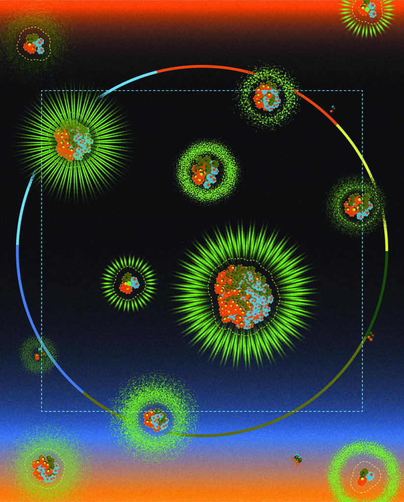
BAD CIRCLES
Long-ignored rings of DNA prove critical in cancer development and drug resistance
Tiny cancer-associated DNA circles rolled into researchers’ awareness in the mid-1960s, when microbiologists first spied the small structures inside cells, bobbing near much larger chromosomes. But their critical role in cancer development and progression began to be appreciated only when advanced DNA sequencing techniques allowed researchers to learn that they frequently contained cancer-associated genes called oncogenes.
The circles, called extrachromosomal DNA, or ecDNA, bounced further into the spotlight in 2022 when professor of pathology Paul Mischel, MD, and his team of international collaborators received a $25 million grant from Cancer Grand Challenges — a research initiative co-founded by Cancer Research UK and the National Cancer Institute to take on some of cancer’s toughest problems.
In November 2024, the team published a trio of landmark papers in Nature describing the biology of ecDNA and a way cancer treatments could go after the circles.
The first of these papers detailed the prevalence and impact of ecDNA in nearly 15,000 people with 39 different types of cancer. Another paper described a cancer treatment targeting the circles that’s already in clinical trials. The third paper revealed the unusual way the circles are passed down during cell division, a mode of transmission that overthrows a fundamental law of genetics — that chromosomes assort randomly between cells during division.
“We’re in the midst of a completely new understanding of a common and aggressive mechanism that drives cancer,” said Mischel, who holds the Fortinet Founders Professorship. “Each paper alone is noteworthy; taken together, they represent a major inflection point in how we view cancer initiation and evolution.”
Howard Chang, MD, PhD, a professor of dermatology and genetics and the Virginia and D.K. Ludwig Professor in Cancer Research, is a close collaborator with Mischel and co-author on all three papers.
“We’re in the midst of a completely new understanding of a common and aggressive mechanism that drives cancer.”
Paul Mischel, professor of pathology and a Stanford University Sarafan ChEM-H institute scholar
The findings of the studies are stunning. The researchers found ecDNA in 17% of the samples and learned that certain tumor types, including breast and bone cancer, were more likely than others to harbor the circles. They also showed that, in addition to oncogenes, ecDNA molecules often harbor genes associated with dampening the immune response to cancer. Late-stage cancers were more likely to have ecDNA, and the presence of the circles correlated with metastasis and poorer overall survival.
The circles’ power stems from their ability to work together. When a circle with a cancer-driving oncogene snuggles next to a circle with a DNA sequence that kicks off the translation of the oncogene into its havoc-wreaking protein, the production of that protein shifts into overdrive.
Cancer cells respond to this flood of growth messages by dividing repeatedly in the growing tumor.
The nefarious cycle doesn’t stop there. EcDNA molecules that work together tend to stay together during cell division, confounding a long-standing understanding of how independent DNA structures are inherited. This increases the odds that a daughter cell will end up with combinations of ecDNA that work well together.
“These kinds of ‘jackpot’ inheritance events can confer a huge growth advantage,” Chang said.
But the insatiable need of the circles to churn out cancer-driving proteins is also a weakness the researchers learned to exploit. Blocking the ability of ecDNA-containing cells to juggle the conflicting processes of gene expression (needed to make proteins) and DNA replication (an essential step in cell division) causes the cells to falter and die, they found.
The researchers’ results have sparked two early phase clinical trials sponsored by Boundless Bio, a San Diego-based oncology company co-founded by Mischel and Chang. The trials are enrolling people with certain types of cancers who have multiple copies of oncogenes or of a gene called MAPK that’s involved in resistance to treatment encoded on ecDNAs.
Mischel is a Stanford University Sarafan ChEM-H institute scholar and a member of the Stanford Cancer Institute. Chang began a leave of absence from Stanford Medicine in December 2024 to serve as senior vice president of research and chief scientific officer at Amgen.
HOW THE MIND CAN
HELP THE BODY
Watching a series of short videos on mindset improved cancer patients’ quality of life
So much about cancer presents as hard, immutable facts. The disease is Stage 3. The tumor is 5 millimeters. The recommended treatment is this many chemotherapy sessions.
But cancer happens to humans, with all their associated baggage, and two patients with the same disease might have wildly different experiences.
“In my years of clinical work, it became obvious that the way different people experienced cancer and treatment depended on many reasons that were not just related to the doses of medications or the stage of disease,” said Lidia Schapira, MD, director of the Stanford Medicine Cancer Survivorship Program. “We didn’t have a rigorous way of understanding those differences.”
Now, Schapira, who’s also a professor of medicine and member of the Stanford Cancer Institute, and her colleagues in oncology, psychology and psychiatry at Stanford are not only studying those differences but developing ways to help patients who have the hardest time coping with cancer. They created a series of short films that they’ve found can improve cancer patients’ mindsets, or the set of attitudes each person holds, which in turn improves how they physically experience their disease and treatment.
‘There’s a lot of nuance’
Call it mind over matter — but don’t call it the power of positive thinking.
“A lot of people assume when we talk about mindsets that that’s just positive thinking,” said Alia Crum, PhD, associate professor of psychology and head of Stanford’s Mind and Body Lab. “We’re not saying you should be happy you have cancer. There’s a lot of nuance in what psychological construct we’re targeting.”
The good news is that even when facing something as serious and uncertain as cancer, mindsets can change. That’s the finding of a study led by Crum and former Stanford University graduate student Sean Zion, PhD, that was published in summer 2023 in Psycho-Oncology.
The team spent several years studying the spectrum of how patients view cancer — from catastrophe to manageable to a growth opportunity (which is actually surprisingly common) — and whether they viewed their bodies as capable or adversarial. The researchers then created six documentary-style films designed to shift patients’ attitudes about their conditions toward the capable and manageable side of the spectrum. (One of the study’s co-authors, Jonathan Berek, MD, the Laurie Kraus Lacob Professor, is both an oncologist and a documentary filmmaker, and he shot and edited the films.)
“Helping people feel like they have mastery and expertise in living with cancer and coping with it is good for them medically as well as psychologically.”
David Spiegel, professor of psychiatry
The films ranged from 4 to 12 minutes long and included interviews with experts as well as cancer survivors and caregivers about their experiences. Each video targeted a certain aspect of mindset, such as how one can come to appreciate how capable their bodies really are as they endure and respond to cancer treatment. Participants were also asked to answer questions reflecting on the video content.
The team recruited 361 recently diagnosed patients with a wide variety of cancer types from across the U.S. Half were asked to watch the videos at certain times over the course of their treatments. Participants who watched the videos reported around an 8% increase in their health-related quality of life as well as less bothersome side effects from treatment, while those who didn’t watch the videos saw a slight decrease in their quality of life.
The team has received funding from the National Institutes of Health to expand their study to more patients and to include biological measurements of stress and inflammation through blood samples. They’re hoping to launch recruitment for this new study in early 2025. Eventually, they want to make the videos widely available to cancer patients.
“Helping people feel like they have mastery and expertise in living with cancer and coping with it is good for them medically as well as psychologically,” said David Spiegel, MD, a co-author of the study, professor of psychiatry and the Jack, Lulu, and Sam Willson Professor in Medicine. “Changing your mind can help to change your body.”
AIMING FOR A
RADIATION BULL’S-EYE
Inventions are on the horizon to destroy cancer with more precision
Ever since X-rays were discovered more than a century ago, radiation has been used to treat cancer. Today, various types of high-energy radiation can be directed at tumors anywhere in the body, and about two-thirds of cancer patients receive radiation therapy at some point in their treatment.
But the central conundrum of radiation therapy remains the same — how to deliver an obliterating dose of radiation to a tumor while causing minimal damage to surrounding tissue. Several new innovations at Stanford Medicine, including real-time tumor tracking, rapid radiation delivery and ultra-compact proton therapy, are closing in on solutions.
These recent advances in radiation therapy are not only creating a renaissance in the field but are also leading to treatments for previously untreatable cancers.
Delivering radiation precisely poses a major challenge because any tumor — simply by residing in a living, breathing body — is a moving target. To ensure that they’re treating the entire tumor, radiation oncologists might resort to irradiating a larger area that includes healthy tissue. To minimize harm to healthy tissue, they might use technology that turns the radiation on only when the tumor moves into a target area. Even then, the targets are mapped by imaging, such as PET scans, typically days or weeks in advance, which means organs might have shifted in the meantime.
But a first-of-its-kind machine at Stanford Medicine can now track tumors in real time. Known as SCINTIX, the machine uses an integrated PET scanner to visualize a tumor during treatment, redirecting the radiation several times a second. “If a patient were to move a little bit during treatment, we would have the peace of mind that this is tracking and following the tumor,” said Lucas Vitzthum, MD, clinical associate professor of radiation oncology.
It’s the difference between a static, paper road map and an onboard GPS system.
“It’s using the biological signal from a tumor to track it and deliver radiation in real time,” Vitzthum said. “It’s really the only thing like it.”
Based on clinical research led by Vitzthum, the FDA approved the technology for lung and bone cancer, and Stanford Medicine has treated four patients using the new system. With further development, including a hardware update planned for early 2026, Vitzthum hopes SCINTIX will be able to track and treat multiple tumors at once.
More than a decade ago, Billy Loo, Jr., MD, PhD, professor of radiation oncology, began pondering another strategy to minimize collateral damage from radiation. Existing strategies were all “compensating for the assumption that it takes longer to give the radiation than for the body to move,” he said. “We started thinking, what if we could turn around that assumption? What if we could develop technology to do the treatment much faster than the motion?”
With colleagues at SLAC National Accelerator Laboratory, Loo’s team helped develop more powerful radiation beams that could deliver the same dose of radiation hundreds of times faster — in a fraction of a second. Soon afterward, researchers at the Curie Institute in France discovered that such ultra-rapid delivery of radiation — known as FLASH — could kill tumors in mice just as effectively with substantially less damage to normal tissue.
“That puts a totally new spin on what we’re trying to do because not only can we make the treatment more precise and freeze the motion, but we also get this additional biological advantage.”
Billy Loo Jr., professor of radiation oncology
“That puts a totally new spin on what we’re trying to do because not only can we make the treatment more precise and freeze the motion, but we also get this additional biological advantage,” Loo said. “That’s really paradigm shifting.”
Loo’s is now one of the leading labs focused on the biological aspects of FLASH. Preclinical results in different animal models and organ systems are promising. In mice, for example, FLASH can blast tumors in the abdomen without damaging stem cells in the intestines and zap tumors in the brain while preserving cognitive function. Loo and SLAC collaborators are working on a new class of linear accelerator that can generate even more powerful and focused FLASH for use in humans.
In the near term, a limited version of FLASH may be possible through proton therapy, an alternative to X-rays that delivers higher doses of radiation more precisely. While X-rays inevitably penetrate beyond a target, protons can be stopped at a specific depth.
“With the right energy, a proton can penetrate deep enough to reach a tumor and actually deposit most of its energy at that depth, which helps you get more dose to where you want it and less to where you don’t want it,” Loo said.
Proton therapy can also be delivered from multiple angles so the highest dose is concentrated at the tumor while only a fraction of the dose goes through healthy tissue. Unfortunately, proton therapy has been far less accessible than traditional radiation therapy with X-rays because it requires much larger accelerators and beamlines, which are typically housed in facilities the size of a football field. More compact versions have shrunk that to the size of a basketball court, but they still need to be three stories high so the machine can rotate around the patient. The closest proton therapy facility to the Bay Area is an eight-hour drive south, in San Diego.
Let’s get smaller
Through a collaboration with two companies, Stanford Medicine is installing the first ultracompact proton therapy machine, which will fit into a normal radiation treatment room. The machine combines a compact linear accelerator with another innovation — rotating the patient instead of the machine. Typically, patients receive radiation treatment lying down, but the new design positions the patient upright on a sort of high-tech lazy Susan.
“It’s a convenient and ergonomic way to rotate the patient rather than rotating a huge machine around the patient,” Loo said. The ultracompact machine is expected to be ready for treating patients by fall 2025.
Loo tells his residents that almost no radiation technique they use today, from the simplest to the most complex treatments, is the same as it was when he was a resident 20 years ago. “That tells you how the field has evolved just with incremental improvements,” he said. “Now, we’re working on potential quantum leaps.”
OPTIMIZING CARE FOR A STUBBORN LUNG CANCER
A med student’s effort to test a treatment for EGFR-mutated lung cancer comes to fruition
The oncologist’s toolbox carries a growing selection of fine-tuned instruments with specialized functions. Known as targeted therapies, these treatments manipulate specific molecules and genes in cancer cells to inhibit their growth or make them more vulnerable to the immune system.
But precision instruments have to be applied precisely. As new targeted therapies emerge, researchers must constantly evaluate how and when to best integrate them into tried-and-true treatment protocols. That means every clinical trial has the potential to create a shift in standard practice.
Lung cancer, in particular, is increasingly defined and treated according to its genetic mutations. About 10% to 15% of lung cancer patients in the United States have a mutation in their epidermal growth factor receptor, or EGFR, a protein on the surface of cells that normally helps control cell growth.
“When EGFR develops a mutation that drives too much cell proliferation, that’s when you develop a tumor,” said Jacqueline Aredo, MD, a clinical fellow in medical oncology.
EGFR-mutated lung cancer disproportionately affects people of Asian background, women, nonsmokers and younger patients. By the time the disease is diagnosed, it is often advanced or has grown too bulky for surgery to be a viable option.
A few years ago, when Aredo was a Stanford medical student, she led a multicenter clinical study evaluating the effect of an immunotherapy drug, durvalumab, on patients with locally advanced EGFR-mutated lung cancer — tumors that had spread to nearby tissues or lymph nodes but not to other organs. The drug was promising enough that the FDA had already approved it for use after chemoradiation to prevent lung cancer from recurring and to improve survival.
But the Stanford Medicine study found that the drug may be doing more harm than good in patients with EGFR-mutated lung cancer: Not only did they receive no benefit from the drug, but they were also likely to suffer serious immune-related side effects.
“Standard of care for these patients had been chemoradiation followed by durvalumab immunotherapy,” said Aredo, who graduated in 2021. “Immunotherapy tends to benefit most patients with lung cancer, but EGFR-mutant lung cancers tend to have a more immune-inert phenotype.”
Aredo was eager to find something else they could offer patients with locally advanced EGFR-mutated lung cancer. Looking over the data from the durvalumab study, she noticed that a few patients in the control group, who did not receive the drug, did remarkably well and were less likely to have cancer recur.
“I saw that the survival curve showed us two different populations. It was a curious finding,” Aredo said. The patients who did better had received a different type of targeted drug — an EGFR inhibitor — at some point in their treatment.
“I saw that the survival curve showed us two different populations. It was a curious finding.”
Jacqueline Aredo, a clinical fellow in medical oncology
“When we found this group of patients who were receiving EGFR inhibitors, either before or after chemoradiation, we thought perhaps that might lead to a better strategy,” said Aredo, who began thinking about a new trial.
In fact, an ongoing trial of an EGFR inhibitor called osimertinib for locally advanced EGFR-mutated lung cancer after chemoradiation was already underway. Based on the promising results of that trial — which found an 84% reduction in the risk of disease recurrence or death — and FDA approval in September 2024, osimertinib has become a standard post-chemoradiation treatment for these patients.
“This showed just how important it is to include a targeted drug like osimertinib into the treatment algorithm for these patients,” Aredo said.
But Aredo thinks there’s room for improvement. She and her mentors are realizing the project she conceived in medical school. They’ve launched an international clinical trial, called NEOLA, to test whether receiving osimertinib before and after chemoradiation benefits patients with locally advanced EGFR-mutated lung cancer — potentially changing standard protocol again. They hope the drug can help shrink tumors ahead of chemoradiation, enhance the treatment and prevent recurrence.
Aredo, who recently returned to Stanford Medicine for her fellowship, helped design the new trial and serves on the international steering committee, an unusual position for a trainee. She credits the encouragement from her mentors, Heather Wakelee, MD, the Winston Chen and Phyllis Huang Professor, and Joel Neal, MD, PhD, professor of medicine. “They’ve supported me through all phases of this journey,” she said. “My experience really shows the tremendous impact that mentorship can have on realizing the potential of medical trainees.”
And she’s keeping an eye on the horizon. There’s increasing evidence that people with early stage lung cancers can also benefit from the addition of targeted therapies to their treatment.
“Because targeted therapies for lung cancer were first established for advanced or metastatic cancers, there wasn’t a need to test for mutations in early stage patients,” Aredo said. That, too, may soon change.
TURNING CANCER DRIVERS INTO CANCER KILLERS
Researchers tether proteins together, causing tumors to self-destruct
Like many of us during the early days of the COVID-19 pandemic, Gerald Crabtree, MD, got in the habit of taking long walks near his house when he couldn’t go to work. Like only some of us, however, the Stanford Medicine cancer biologist had a eureka moment during one such walk.
The idea that struck him was for a new kind of cancer therapy marrying two major milestones in biology: the discovery that certain mutated genes, known as oncogenes, drive most forms of cancer and the finding that multicellular creatures undergo large swaths of programmed cell death, known as apoptosis, for the greater good of the organism. Our bodies get rid of some 60 billion cells every day through apoptosis, and it’s precise enough that typically no cell dies that should remain alive.
“I thought, this is the kind of mechanism we should bring to cancer treatment,” said Crabtree, the David Korn, MD, Professor in Pathology and a member of the Stanford Cancer Institute. “The trick was to get the cancer driver to activate these cell-death pathways.”
Crabtree partnered with Nathanael Gray, PhD, the Krishnan-Shah Family Professor and professor of chemical and systems biology at Stanford Medicine, to figure out how to merge these two cellular processes. They settled on a technique known as chemical inducers of proximity, in which small molecules bring proteins together, allowing them to act on each other in ways they normally wouldn’t.
The team first turned to the blood cancer known as diffuse large B-cell lymphoma, which is driven by an oncogene known as BCL6. In this type of cancer, the BCL6 protein sits on apoptosis genes, keeping them switched off and allowing the cancer cells to stay immortal. To flip the script on BCL6’s action, the researchers developed a molecular “glue” that links the protein to other proteins that activate genes.
“We’re taking a cancer cell’s survival factor and turning it into a death factor,” said Gray, who is also a member of the cancer institute. “This compound is giving us a new kind of pharmacology that we wouldn’t have necessarily anticipated from either protein alone.”
So far, they’ve made two such “unnatural unions,” as Gray calls them, with the help of linking molecules (the “glue”): one pairing BCL6 to a protein known as CDK9, which they described in a study published in Science last year, and another pairing BCL6 with a protein called BRD4, detailed in a 2023 paper in Nature.
“We’re taking a cancer cell’s survival factor and turning it into a death factor.”
Nathanael Gray, chemical and systems biology professor
In both cases, they showed that the chimeric proteins act to turn on apoptosis genes where they are normally shut off, ultimately killing the cancer cells. Because BCL6 sits on more than a dozen independent cell death genes, using this protein to switch on the genes appears to activate a robust and highly specific process of cell killing.
In their most recent paper, the researchers tested the BCL6-CDK9 linking molecule in dozens of different types of cancer cells in the lab. They found it precipitated the death of only diffuse large B-cell lymphoma tissue and did not harm the other kinds of cancer cells. The linking molecule doesn’t activate the killing of cells that don’t carry BCL6, which is not produced in other kinds of cancer, the study found.
In the future, the researchers hope to build molecules targeting oncogenes that are widespread across different tumor types. They’ve also tested the linking molecule in healthy mice and found no indication of toxicity, even though the molecule also led to the death of some of the animals’ healthy immune cells.
They’re now testing the linking molecules in mice with lymphoma. Through work conducted at Shenandoah Therapeutics, a biotech startup they co-founded, Crabtree and Gray hope to gather enough preclinical data to move the technique to clinical trials.