Vicious circles
Cancer’s deadly weapon – rings of DNA – have been hiding in plain sight
Location, location, location. It’s not just important in real estate but also in biology and, apparently, research seminars.
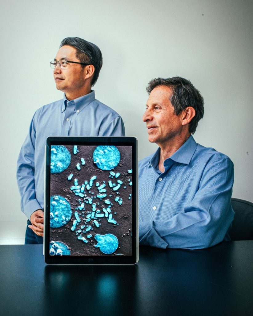
It was a Tuesday afternoon in December 2017, and Paul Mischel, MD, then a cancer biologist at UC San Diego, had just finished giving a talk at Stanford describing a surprising observation: small circles of DNA in cancer cells bobbing in the cells’ nuclei, untethered to nearby chromosomes — the multiple long chains of DNA that comprise the cells’ genetic material.
The circles, known as extrachromosomal DNA, or ecDNA, had been dismissed for decades by mainstream geneticists as a biological fluke. But a few years earlier, Mischel had begun to suspect there was more to the free-floating, SpaghettiOs-shaped structures.
His hunch was right. We now know that the circles, which are only occasionally found in healthy cells, are chockablock with cancer-causing genes. They are a primary driver in cancer growth and the evolution that helps some tumors evade drug therapies within weeks or months. Unfortunately, they are not rare: 1 in 3 cancer patients, often those with the most aggressive types of cancer, have high levels of ecDNA in their tumor cells.
Recently, the circles’ importance has been internationally recognized. In 2021, the National Cancer Institute and Cancer Research UK partnered to select ecDNA as one of eight Cancer Grand Challenges with the potential to advance cancer research and improve the lives of people with cancer.
And in June, Mischel and his team were selected from a panel of global applicants to receive $25 million from the partnership to continue their research into ecDNA in cancer.
“We now have an unparalleled opportunity to move from incremental research advances to transformational science,” Mischel said. “Patients whose cancer cells have lots of ecDNA fare much more poorly than their peers do. There is a massive medical need to understand how they function.”
But how do you get to the bottom of a circle?
“After the talk, Howard came up and said, ‘Hey Paul, I think we might be seeing something similar in our data. It was really a life-changing moment for me.”
Paul Mischel, MD
At the time of the 2017 seminar, only a few researchers were exploring the role of ecDNA in cancer. But Mischel’s audience, including Howard Chang, MD, PhD, a professor of genetics and Stanford Medicine’s Virginia and D.K. Ludwig Professor in Cancer Research, was intrigued. Chang was studying when and how genes are turned on, or expressed, in cancerous and healthy cells.
“After the talk, Howard came up and said, ‘Hey Paul, I think we might be seeing something similar in our data,’” Mischel recalled. “It was really a life-changing moment for me.”
Mischel, who joined Stanford Medicine in 2021 as a professor of pathology, and Chang decided that day to team up to learn more about ecDNA and how it functions in cancer patients. Their results turned traditional genetics on its head and spawned an entirely new field of research.
“The ways in which these circles interact to affect gene expression to drive cancer growth is an entirely new concept in molecular biology,” Chang said. “We believe it will rewrite biology textbooks.”
Chang describes the circles as vicious gangs that terrorize the chromosome-bound genome by ignoring all the understood rules of biology, making cancer therapies for some patients a game of whack-a-mole as tumors evolve drug resistance within days or weeks.
In short, they are agents of chaos. And stopping them has become a primary goal of cancer researchers worldwide.
Now wait a gosh darn minute
Mischel’s talk wasn’t the first time ecDNA had been described in cancers. Microbiologists in the mid-1960s who observed the ring-shaped structures near chromosomes called them “minutes” (with a long vowel i), meaning tiny. Two circles linked together in a figure eight structure were called “double minutes.”
Having named them, but without having the tools to study them in greater detail, biologists for the most part ignored them. Instead, genome biologists focused on mapping the locations of and, later, sequencing individual genes on each of the 23 pairs of chromosomes in each mammalian cell.
Chromosomes are made up of genes and regulatory regions — switches that determine when and where the genes turn on and off — linked arm in arm like the setup for the childhood game of Red Rover.
To fit inside the cramped space of the nucleus, the strand of tens of millions of genetic building blocks twists tightly around itself and winds around packaging proteins called histones, like a line of excited hand-holding kindergartners crowding around a puppy.
“Darwin taught us that genetic variation is the fuel for natural selection. What we were seeing was cancer evolution on steroids. It’s a whole different level.”
Paul Mischel, MD
Until about 50 years ago, geneticists and biologists believed that mammalian cells had two, and only two, copies of each gene — one on each member of the chromosomal pair (with the exception of genes found on the sex chromosomes, which differ in their gene makeup). They also believed chromosomes were aloof: Regulatory regions on one chromosome didn’t affect genes on another.
In the 1970s, however, the late Stanford biologist Robert Schimke, MD, and his lab performed a series of experiments that showed that mammalian cells could in fact harbor more than two copies of certain genes — a concept termed gene amplification.
Importantly, the number of copies of an amplified gene in each cell correlated with the number of minutes or double minutes it had, and the presence of the circles was a key factor in the cells’ ability to rapidly evolve resistance to a common chemotherapy drug.
Although gene amplification in mammalian cells was eventually accepted, Schimke’s first public discussion of the possibility was met with skepticism. He described a session at a Cold Spring Harbor Laboratory symposium in June 1977 in which he broached the possibility as “memorable and stormy.”
Rise of the circles
Schimke’s discovery of gene amplification opened a new era in cancer biology. But despite his identification of ecDNA as one way cells could accumulate more than two copies of a gene, most research focused on a subsequent discovery that gene amplification could also occur on the chromosomes themselves. Curiosity about the circles subsided once again.
One reason for this is that the DNA sequencing technologies first launched in the late 1970s had no way of differentiating DNA on chromosomes from DNA in ecDNA. Also, it was believed (wrongly, it turns out) that ecDNA was rare — occurring in less than 2% of cancers.
But in 2017, Mischel and his colleagues showed that ecDNA is widespread and likely to play a major role in many human cancers.
The roots of the discovery arose from research Mischel and a colleague at UC San Diego were conducting on tumor cells from patients with glioblastoma — a highly aggressive form of brain cancer. Patients with the condition have an amplification of a gene called epidermal growth factor receptor, or EGFR, that encourages cancer cells to divide uncontrollably. At first, the researchers assumed that the extra copies of the gene resided on chromosome 7, near the original gene.
A drug targeting the EGFR protein could be expected to kill the cancer cells and slow tumor growth. But something unexpected happened. Instead of slowing tumor growth, the drug caused the cancer cells to quickly lose extra copies of the gene — much more quickly, in fact, than could be explained with traditional genetics.
Furthermore, when the researchers grew the patients’ cancer cells in the laboratory, each original cell from the tumor gave rise to a colony of cells that included cells with high, low or no copies of the EGFR gene, regardless of the status of the founding cell.
This was also unexpected in the chromosomal-centric view; usually a cell carrying just a few of these genes would give rise only to cells that similarly carry just a few.
“To understand what was going on, we looked inside the nucleus of the cells and got a shock,” Mischel said. “We found that multiple copies of the EGFR gene were located on the ecDNA, rather than on the cells’ chromosomes.”
Unlike chromosomes, which are equitably distributed between daughter cells during cell division, ecDNA swirls unpredictably in the nucleus like bubbles in a bathtub and are portioned out willy-nilly to the daughters. As a result, changes in the overall genetic makeup of the tumor can happen very quickly. Within one or two generations a tumor includes cells that have many, few or none of the circles.
“The regulatory regions on the circles talk in a promiscuous way with other circles, so that switches from completely different chromosomes can affect the expression of genes to which they would never normally have access.”
Howard Chang, MD, PhD
Imagine if each of Darwin’s finches could quickly hatch hundreds of offspring with a nearly infinite variety of beak shapes, without the need to laboriously accumulate random, potentially beneficial mutations along their chromosomes. Now toss this plethora of progeny a nutritious but hard-to-crack peanut, or create a trap that ensnares only the stubby-beaked siblings. No one approach will benefit or harm all flock members simultaneously — with each challenge, some will die and others will thrive.
This is what cancer doctors and drug designers are up against when treating patients whose cancers have high levels of ecDNA, Mischel’s research showed. Like the fanciful finch flock, many aggressive cancers can quickly become resistant to drug therapies targeting cells with high levels of cancer-associated proteins — they simply let those cells die and pivot to others already waiting in the wings that aren’t targeted by the drug.
It’s a bit diabolical.
“Darwin taught us that genetic variation is the fuel for natural selection,” Mischel said. “What we were seeing was cancer evolution on steroids. It’s a whole different level.”
Mischel and his colleagues published their groundbreaking results in Science in 2014, but they were met with “a colossal scratching of heads” and not a little skepticism.
They had only studied cells from glioblastomas — what if the phenomenon were specific to only that cell type?
To find out, the researchers obtained tissue samples from 17 different types of human cancer, including brain, breast, colon, lung and blood cancers. They found that nearly half of all human cancers contain ecDNA, and that these circles frequently carried multiple copies of cancer-driving oncogenes.
The number of circles in each cancer cell varied widely, but they were rarely found in healthy tissue. They published their results in 2017, shortly before the seminar at Stanford.
“We believe that ecDNA is responsible for resistance to so many of our current cancer therapies,” Mischel said. “These circles are driving the production of massive amounts of cancer-promoting proteins.”
Chromosome conundrum
About the same time that Mischel and his colleagues were peering into the nuclei of glioblastoma cells, Chang was grappling with his own mystery as he tried to learn how the three-dimensional structure of chromosomes affects gene expression and cell growth in cancer cells.
Normally tightly wound (remember those excited kindergartners?), the section of the chromosome containing a gene to be expressed must be unpacked so it can be accessed by the relatively bulky protein machines that trundle across genes to copy their sequences into mRNA.
Chang and Stanford colleague and professor of genetics Will Greenleaf, PhD, developed a way to generate a global “accessibility profile” to identify regions of active gene expression.
There was just one problem: The data was indicating there were far too many accessible regions around oncogenes in cancer cells than could be explained by the numbers of known gene switches on chromosomes.
“Most human DNA is not easily accessible,” Chang said. “But these oncogenes were generating such dominant signals in our assays that we had to set them aside to analyze later. It turns out that these oncogenes were on ecDNA.”
He brought up the problem to Mischel after the seminar.
“When Howard showed me the data, I almost fell out of my chair,” Mischel said. Working together, the researchers began to churn out research papers that outlined exactly how the ecDNA circles helped cancer cells thrive.
In 2019, Chang and Mischel published a paper in Nature showing that the oncogenes on the ecDNA are much more active than their chromosomal counterparts.
The researchers soon learned that the shape of the ecDNA molecules was key. “Think about the architecture of a circle,” Mischel said. “On a chromosome, control elements are usually located near the genes they control. But when you take that linear structure and make it into a circle, everything is that much closer. It fundamentally changes how gene regulation happens.”
“The ways in which these circles interact to affect gene expression to drive cancer growth is an entirely new concept in molecular biology. We believe it will rewrite biology textbooks.”
Howard Chang, MD, PhD
Once again, location is important.
“Most of the time we see all the ecDNAs in a cancer cell congregating in a single spot in the nucleus,” Chang said. “We found they form a new structure we called a hub in the nucleus, with multiple oncogenes on tens or hundreds of circles. The regulatory regions on the circles talk in a promiscuous way with other circles, so that switches from completely different chromosomes can affect the expression of genes to which they would never normally have access.”
Think of the Red Rover analogy. It’s possible for whispered instructions to be shared between one child and another further down in the line by creating a loop in the kid “chromosome.” But it’s much more difficult for a child in one chain to get close enough to communicate that same secret message to another in the opposite lineup across the field.
Now imagine the same hand-holding children playing Ring Around the Rosie, spinning and jumping in multiple small groups on a shared playground. It’s child’s play (literally!) for participants in different circles to bump into one another and share confidences.
“This is a really surprising result,” Chang said. “These circles are acting as a collective in ways that biologists have never seen before. The dogma that regulatory regions act only on nearby genes on the same chromosome simply ceases to exist.”
Further research by Chang and Mischel showed that blocking the formation of the protein complex that glues the ecDNAs together causes the circles to fly apart and stops the expression of the oncogenes. In December 2021, they published a bombshell paper in Nature detailing their results and speculating that drugs targeting the hub complex could usher in a new class of therapy for many kinds of cancers.
Full circle
The prospect of eliminating the oncogene expression of all the ecDNAs in a cancer cell in one fell swoop is tantalizing. Chang and Mischel are among several co-founders of a company, Boundless Bio Inc., focused on developing ecDNA-based therapies for patients with intractable cancers.
“I’m most excited about the concept of new therapeutics,” Chang said. “Cancer is a disease of genes. Until now we’ve focused on developing a bespoke solution for each cancer type. If there’s an abnormal enzyme, we try to block that enzyme. But if we can disrupt the hub that binds these circles together, we can potentially treat many types of cancer with one approach.”
Stopping them from forming at all is another possible option. But many questions remain.
“There is so much more we want to learn about ecDNA,” Mischel said. “How do they form, and how are they maintained? Is it possible to stop them from developing? How does the hub work to hold the circles together? And finally, how do they escape the immune system?”
The continuous, rapid selection ecDNAs undergo after each cell division also serves as a built-in research tool to identify which oncogenes are most important in specific types of cancer. “If an ecDNA provides no growth advantage, it would be lost from the cell population right away,” Chang said. “Thus, ecDNAs in cancers tell scientists and physicians exactly what is fueling cell growth.”
Fundamental changes aren’t always easy, or popular. But Mischel and Chang are confident that their findings will lead to a sea change in our understanding of cancer and how to treat it.
“People are visual learners,” Mischel said. “But the molecular world is too small for us to see. So we make maps to help us understand. In cancer genetics, the map has been centered on the role of chromosomes. But these findings have upended this map. Now we are redrawing our map and recharting our course to help patients with the most aggressive forms of cancer.”
More on cancer